. 2010 Mar 4:9:49.
doi: 10.1186/1476-4598-9-49.

Histone deacetylase inhibitor vorinostat suppresses the growth of uterine sarcomas in vitro and in vivo

Affiliations

Histone deacetylase inhibitor vorinostat suppresses the growth of uterine sarcomas in vitro and in vivo

Andelko Hrzenjak et al. Mol Cancer. .

Abstract

Background: Uterine sarcomas are very rare malignancies with no approved chemotherapy protocols. Histone deacetylase (HDAC) inhibitors belong to the most promising groups of compounds for molecular targeting therapy. Here, we described the antitumor effects of suberoylanilide hydroxamic acid (SAHA; vorinostat) on MES-SA uterine sarcoma cells in vitro and in vivo. We investigated effects of vorinostat on growth and colony forming ability by using uterine sarcoma MES-SA cells. We analyzed the influence of vorinostat on expression of different HDACs, p21(WAF1) and activation of apoptosis. Finally, we examined the antitumor effects of vorinostat on uterine sarcoma in vivo.

Results: Vorinostat efficiently suppressed MES-SA cell growth at a low dosage (3 microM) already after 24 hours treatment. Decrease of cell survival was even more pronounced after prolonged treatment and reached 9% and 2% after 48 and 72 hours of treatment, respectively. Colony forming capability of MES-SA cells treated with 3 microM vorinostat for 24 and 48 hours was significantly diminished and blocked after 72 hours. HDACs class I (HDAC2 and 3) as well as class II (HDAC7) were preferentially affected by this treatment. Vorinostat significantly increased p21(WAF1) expression and apoptosis. Nude mice injected with 5 x 10⁶ MES-SA cells were treated for 21 days with vorinostat (50 mg/kg/day) and, in comparison to placebo group, a tumor growth reduction of more than 50% was observed. Results obtained by light- and electron-microscopy suggested pronounced activation of apoptosis in tumors isolated from vorinostat-treated mice.

Conclusions: Our data strongly indicate the high therapeutic potential of vorinostat in uterine sarcomas.


Figures

Figure 1
Figure 1
Vorinostat decreases the growth and colony forming ability of MES-SA cells in vitro. (a) Dose response curve for MES-SA cells treated with five different vorinostat concentrations over three time periods. Cell proliferation was measured by using [3H]thymidine uptake assay. Results are expressed as percentage inhibition of [3H]thymidine incorporation upon vorinostat treatment and normalized to untreated cells. (b) MES-SA cells were treated with 3 μM vorinostat for 24, 48 and 72 hours. Vorinostat induced a strong decrement in the number of treated cells in comparison to untreated control cells. (c) The ability of colony formation was drastically reduced in vorinostat-treated MES-SA cells already after 24 hours and continued to decrease in a time-dependent manner.
Figure 2
Figure 2
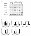
Expression of different histone deacetylases and p21WAF1 in untreated and vorinostat-treated MES-SA cells. (a) Immunoblot analyses were used to examine the expression of different HDACs from class I (HDAC 1, 2 and 3) and class II (HDAC7). Note that not all HDACs were equally affected by vorinostat. HDAC2, 3 and 7 were highly deregulated, whereas HDAC1 was not affected. The expression of cyclin-dependent kinase inhibitor p21WAF1 was highly increased in vorinostat-treated MES-SA cells. (b) Western blot data were quantified densitometrically and beta-tubulin was used as a loading control.
Figure 3
Figure 3
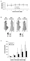
Vorinostat suppressed tumor growth in nude mice xenografts. 5 × 10⁶ MES-SA cells were injected subcutaneously in Nude-Foxn1nu/nu mice (n = 14) and four days later mice were injected either placebo (HOP-β-CD) or 50 mg/kg/day vorinostat dissolved in HOP-β-CD for a total of 21 days. (a) The mice weight curve was stable during the whole treatment indicating that no cachexy was induced by tumor growth. (b) Two representative xenograft mice from each placebo and vorinostat-treated group are shown. Tumors were isolated and tumor weight and volume were determined. In most cases tumors were monolobular, whereas only in two mice two tumor nodules were found at injection side. Obvious differences in tumor volumes were observed between the placebo (2304.7 mm3) versus the vorinostat-treated group (1135.4 mm3). (c) These differences were statistically highly significant at the end of treatment (P = 0.044).
Figure 4
Figure 4
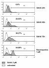
Vorinostat induces apoptosis in MES-SA cells. FACS analysis of MES-SA cells labelled with cleaved caspase-3 antibody conjugated with Alexa Fluor® 488 showed an increased percentage of apoptotic cells in vorinostat-treated cells, correlating with treatment duration. MES-SA cells treated with 1 μM staurosporine for 4 hours were used as a positive control. For numerical analysis two independent experiments, each containing duplicates, were performed. Representative data are shown.
Figure 5
Figure 5
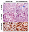
H&E and Ki-67 immunostaining of tumor tissue from nude mice xenografts. (a, c) Solid aggregates of highly atypical tumor cells showing large, round nuclei, prominent nucleoli and abundant cytoplasm were visible in tumors isolated from the placebo group. (b) In vorinostat-treated tumors, highly atypical tumor cells associated with abundant apoptosis were seen. Areas of apoptosis (marked by arrows) show tumor cells with smaller, dark nuclei with condensed chromatin and small rim of cytoplasm. Note the presence of vacuolated cytoplasm, fragmented tumor cells and isolated nuclei of tumor cells. (d) Aggregate of tumor cells with an apoptotic body characterized by dense eosinophilic cytoplasm and hyperchromatic nuclear fragments (marked by arrow). (e, f) More than 95% of tumor cells showed a positive nuclear immunoreaction for Ki-67, both in placebo and in vorinostat-treated tumors.
Figure 6
Figure 6
Representative transmission electron microscopy of tumor tissue isolated from vorinostat-treated mouse xenografts. (a) Shrunken nuclei with condensed and fragmented chromatin (arrow heads) in comparison to non-apoptotic nuclei (arrows). (b) Micrograph of the boxed area of panel A at higher magnification.

References

    1. Harker WG, MacKintosh FR, Sikic BI. Development and characterization of a human sarcoma cell line, MES-SA, sensitive to multiple drugs. Cancer Res. 1983;43:4943–4950. - PubMed
    1. Sleijfer S, Seynaeve C, Verweij J. Gynaecological sarcomas. Curr Opin Onco. 2007;19:492–496. doi: 10.1097/CCO.0b013e3282748eaa. - DOI - PubMed
    1. Gunawan B, Braun S, Cortes MJ, Bergmann F, Karl C, Füzesi L. Characterization of a newly established endometrial stromal sarcoma cell line. Int J Cancer. 1998;77:424–428. doi: 10.1002/(SICI)1097-0215(19980729)77:3<424::AID-IJC19>3.0.CO;2-7. - DOI - PubMed
    1. Kakuno Y, Yamada T, Mori H, Narabayashi I. Establishment and characterization of a cell line (OMC-9) originating from a human endometrial stromal sarcoma. Human Cell. 2008;21:46–55. doi: 10.1111/j.1749-0774.2008.00050.x. - DOI - PubMed
    1. Marks PA, Richon VM, Miller T, Kelly WK. Histone deacetylase inhibitors. Adv Cancer Res. 2004;91:137–168. full_text. - PubMed

Publication types

MeSH terms