Review
. 2010 May-Jun;1799(5-6):353-64.
doi: 10.1016/j.bbagrm.2010.02.005. Epub 2010 Mar 2.

Modulation of transcription factor function by O-GlcNAc modification

Affiliations
Review

Modulation of transcription factor function by O-GlcNAc modification

Sabire Ozcan et al. Biochim Biophys Acta. 2010 May-Jun.

Abstract

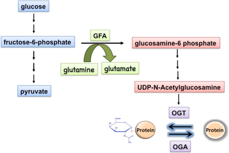
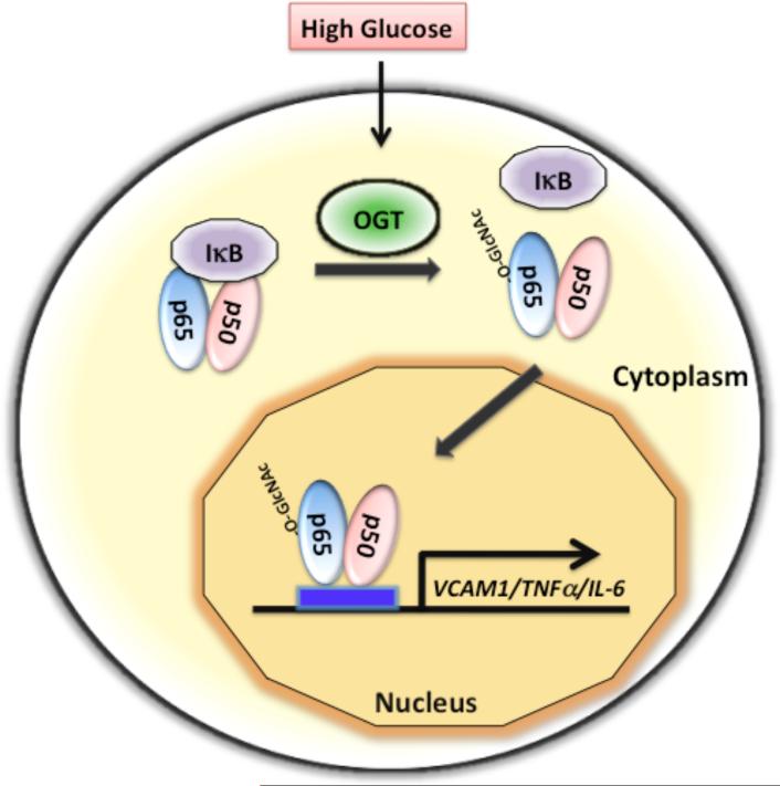
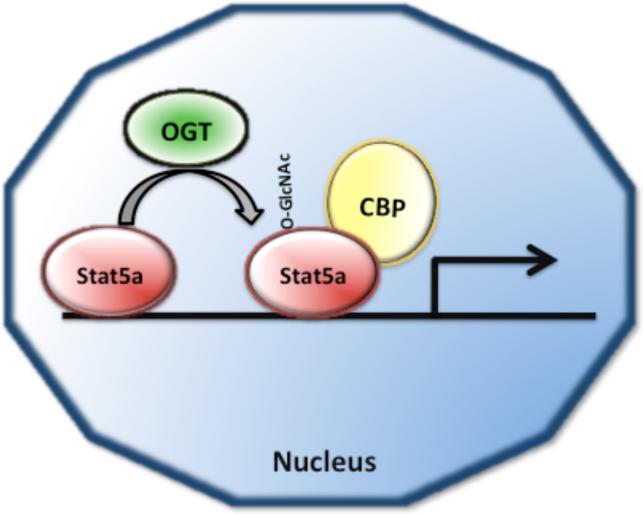
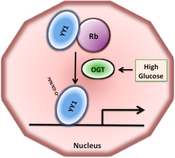
O-linked beta-N-acetylglucosamine (O-GlcNAc) modification of nuclear and cytoplasmic proteins is important for many cellular processes, and the number of proteins that contain this modification is steadily increasing. This modification is dynamic and reversible, and in some cases competes for phosphorylation of the same residues. O-GlcNAc modification of proteins is regulated by cell cycle, nutrient metabolism, and other extracellular signals. Compared to protein phosphorylation, which is mediated by a large number of kinases, O-GlcNAc modification is catalyzed only by one enzyme called O-linked N-acetylglucosaminyl transferase or OGT. Removal of O-GlcNAc from proteins is catalyzed by the enzyme beta-N-acetylglucosaminidase (O-GlcNAcase or OGA). Altered O-linked GlcNAc modification levels contribute to the establishment of many diseases, such as cancer, diabetes, cardiovascular disease, and neurodegeneration. Many transcription factors have been shown to be modified by O-linked GlcNAc modification, which can influence their transcriptional activity, DNA binding, localization, stability, and interaction with other co-factors. This review focuses on modulation of transcription factor function by O-linked GlcNAc modification.


Figures

Fig. 1
Fig. 1. O-GlcNAc modification is linked to glycolysis via the hexosamine biosynthetic pathway (HBP)
Only a small fraction of the glucose (2-5%) enters the HBP, which starts with the conversion of the glycolytic metabolite fructose-6-phosphate and glutamine to glucosamine-6-phosphate and glutamate by the rate-limiting enzyme GFA. The end product of HBP is UDP-N-acetylglucosamine, which is used by OGT as substrate to modify proteins by O-GlcNAc linkages. This modification is reversible, and proteins are deglycosylated by the O-GlcNAcase (OGA).
Fig. 2
Fig. 2. High glucose mediated O-GlcNAc modification of NF-κB disrupts its interaction with the inhibitor IκB and causes nuclear translocation of NF-κB
The transcription factor NF-κB, consisting of two subunits (p50 and p65), is normally sequestered in the cytoplasm by its interaction with the inhibitor IκB. O-linked GlcNAc modification of NF-κB p65 subunit in response to high glucose disrupts its interaction with IκB, leading to nuclear accumulation and activation of NF-κB target genes, such as VCAM-1, TNFα, and IL-6.
Fig. 3
Fig. 3. O-GlcNAc modification of Stat5a enhances its association with the co-activator CREB binding protein CBP
Posttranslational modification of Stat5a with O-GlcNAc has been proposed to enhance transactivation of Stat5a dependent gene expression by promoting the association of Stat5a with the transcriptional co-activator CBP.
Fig. 4
Fig. 4. The interaction of YY1 with Rb is disrupted by O-GlcNAc modification of YY1
Association of YY1 with the retinoblastoma protein Rb inhibits YY1 binding to DNA. O-GlcNAc modification of YY1 increases upon exposure to high glucose and disrupts YY1 interaction with Rb, increasing the ability of YY1 to bind to DNA and to activate transcription.
Fig. 5
Fig. 5. The stability of the tumor suppressor protein p53 is regulated by O-GlcNAc modification
In unstressed cells, p53 is phosphorylated by CSN (COP9 signalosome)-associated kinases, and phosphorylated p53 interacts with the ubiquitin ligase Mdm2, which promotes the proteasomal degradation of p53. In stressed cells, O-GlcNAc modification of p53 prevents its degradation by the proteasome.
Fig. 6
Fig. 6. The function of the insulin gene transcription factors Pdx-1, NeuroD1, and MafA is regulated by O-GlcNAc modification
Activation of insulin gene transcription in pancreatic beta cells requires the synergistic interaction of the transcription factors Pdx-1, NeuroD1, and MafA. O-GlcNAc modification of Pdx-1 enhances the DNA binding capability of Pdx-1. NeuroD1 is normally localized to the cytoplasm, but translocates into the nucleus in response to high glucose mediated by O-GlcNAc modification. MafA expression is induced by high glucose and requires O-GlcNAc modification of an unknown transcriptional regulator.
Fig. 7
Fig. 7. O-GlcNAc modification regulates gluconeogenic gene expression in liver during diabetic conditions
Chronic hyperglycemia or diabetes causes induction of gluconeogenic gene expression by O-GlcNAc modification of the transcription factors CREB and FoxO-1 and their co-activators CRTC2 and PGC-1α, respectively. O-GlcNAc modification of CRTC2 under diabetic conditions causes CRTC2 translocation into the nucleus where it interacts with CREB to activate gluconeogenic gene expression. The co-activator PGC-1α has been demonstrated to be O-GlcNAc modified and to recruit OGT to FoxO-1, promoting O-GlcNAc modification of FoxO-1 and activation of gluconeogenic genes.
Fig. 8
Fig. 8. Modulation of Sp1 function by O-GlcNAc modification involves multiple mechanisms
Modification of Sp1 with O-GlcNAc has been shown to cause its nuclear localization thereby leading to enhanced transcription. O-GlcNAc modification can also lead to transcriptional repression by Sp1 by recruitment of the co-repressor mSin3A via OGT. There is also evidence that O-GlcNAc modification of Sp1 disrupts its interaction with the TATA binding protein-associated factor TAF110.

Similar articles

Cited by

References

    1. Torres CR, Hart GW. Topography and polypeptide distribution of terminal N-acetylglucosamine residues on the surfaces of intact lymphocytes. Evidence for O-linked GlcNAc. J Biol Chem. 259(1984):3308–3317. - PubMed
    1. Davis LI, Blobel G. Identification and characterization of a nuclear pore complex protein. Cell. 45(1986):699–709. - PubMed
    1. Holt GD, Hart GW. The subcellular distribution of terminal N-acetylglucosamine moieties. Localization of a novel protein-saccharide linkage, O-linked GlcNAc. J Biol Chem. 261(1986):8049–8057. - PubMed
    1. Holt GD, Snow CM, Senior A, Haltiwanger RS, Gerace L, Hart GW. Nuclear pore complex glycoproteins contain cytoplasmically disposed O-linked N-acetylglucosamine. J Cell Biol. 104(1987):1157–1164. - PMC - PubMed
    1. Shafi R, Iyer SP, Ellies LG, O'Donnell N, Marek KW, Chui D, Hart GW, Marth JD. The O-GlcNAc transferase gene resides on the X chromosome and is essential for embryonic stem cell viability and mouse ontogeny. Proc Natl Acad Sci U S A. 97(2000):5735–5739. - PMC - PubMed

Publication types

MeSH terms