

Dynamic loading enhances integrative meniscal repair in the presence of interleukin-1
- PMID: 20202487
- PMCID: PMC2872683
- DOI: 10.1016/j.joca.2010.02.009
Dynamic loading enhances integrative meniscal repair in the presence of interleukin-1
Abstract
Objective: Meniscal tears are a common knee injury and increased levels of interleukin-1 (IL-1) have been measured in injured and degenerated joints. Studies have shown that IL-1 decreases the shear strength, cell accumulation, and tissue formation in meniscal repair interfaces. While mechanical stress and IL-1 modulate meniscal biosynthesis and degradation, the effects of dynamic loading on meniscal repair are unknown. The purpose of this study was to determine the effects of mechanical compression on meniscal repair under normal and inflammatory conditions.
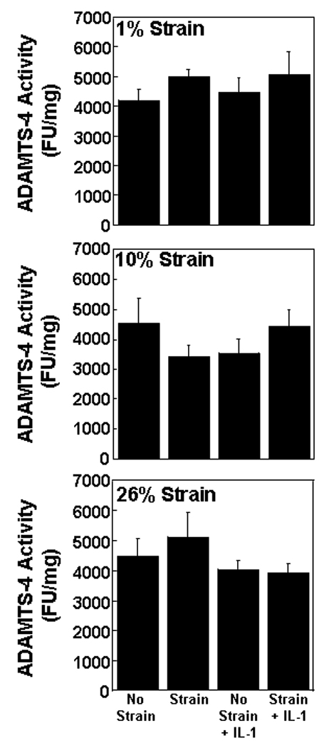
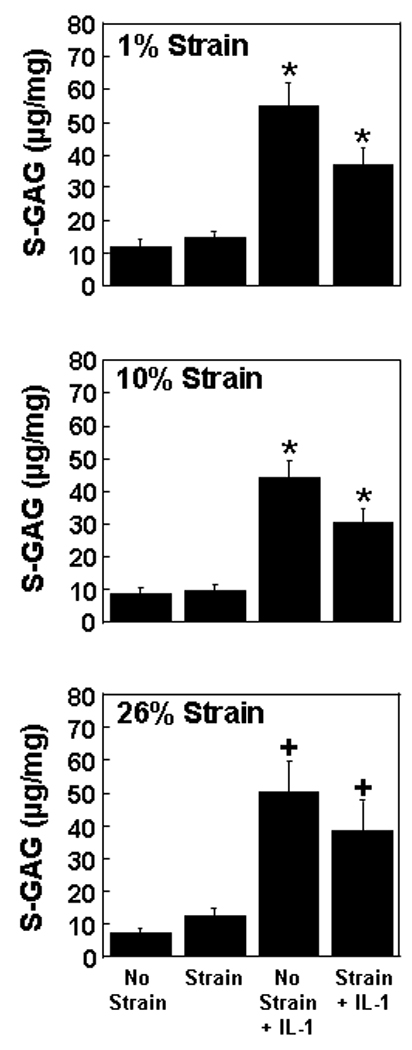
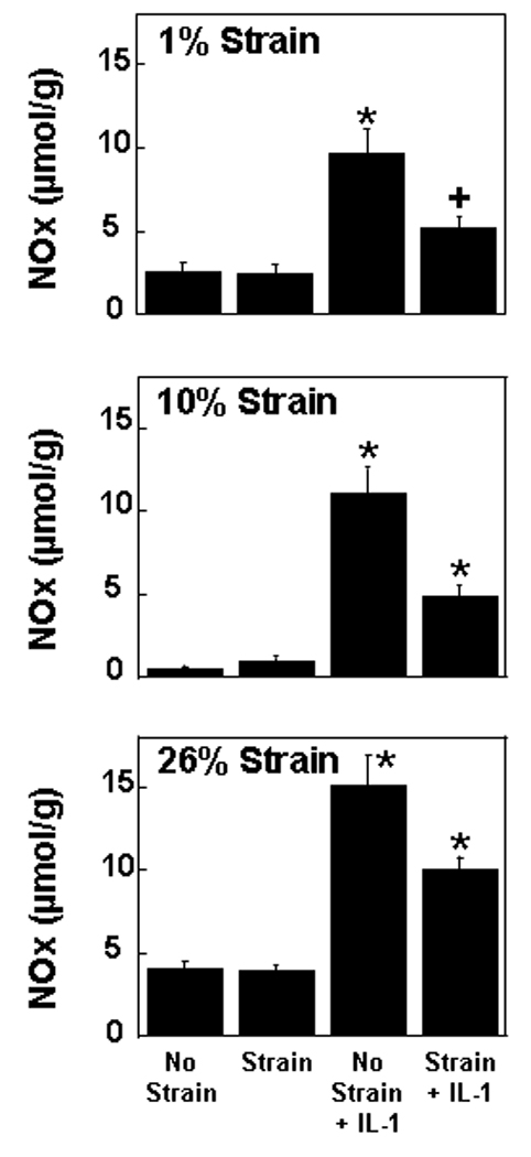
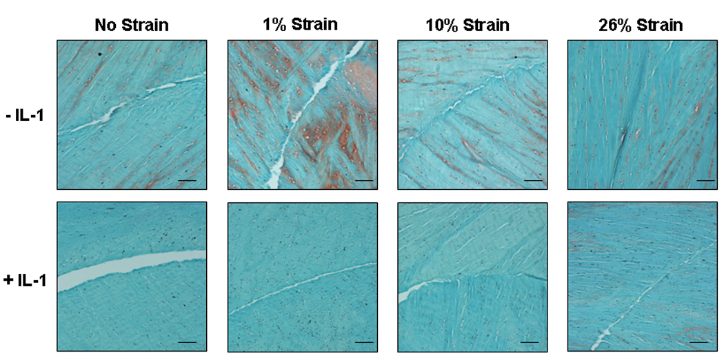
Experimental design: Explants were harvested from porcine medial menisci. To simulate a full-thickness defect, a central core was removed and reinserted. Explants were loaded for 4h/day at 1 Hz and 0%-26% strain for 14 days in the presence of 0 or 100 pg/mL of IL-1. Media were assessed for matrix metalloproteinase (MMP) activity, aggrecanase activity, sulfated glycosaminoglycan (S-GAG) release, and nitric oxide (NO) production. After 14 days, biomechanical testing and histological analyses were performed.
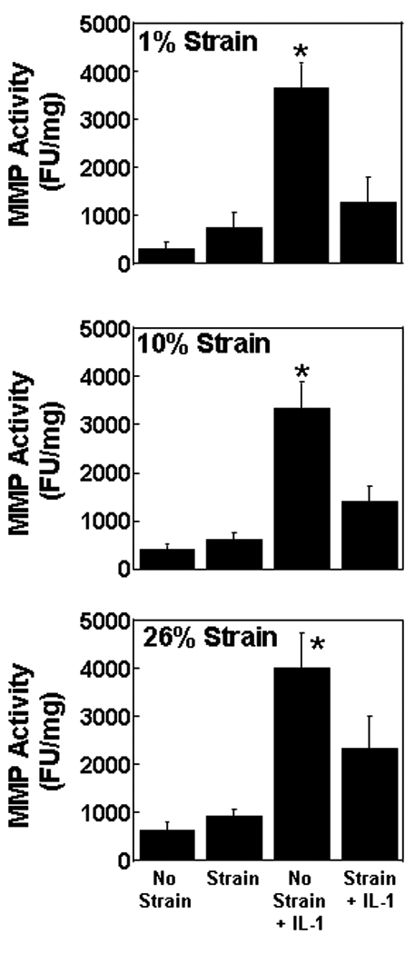
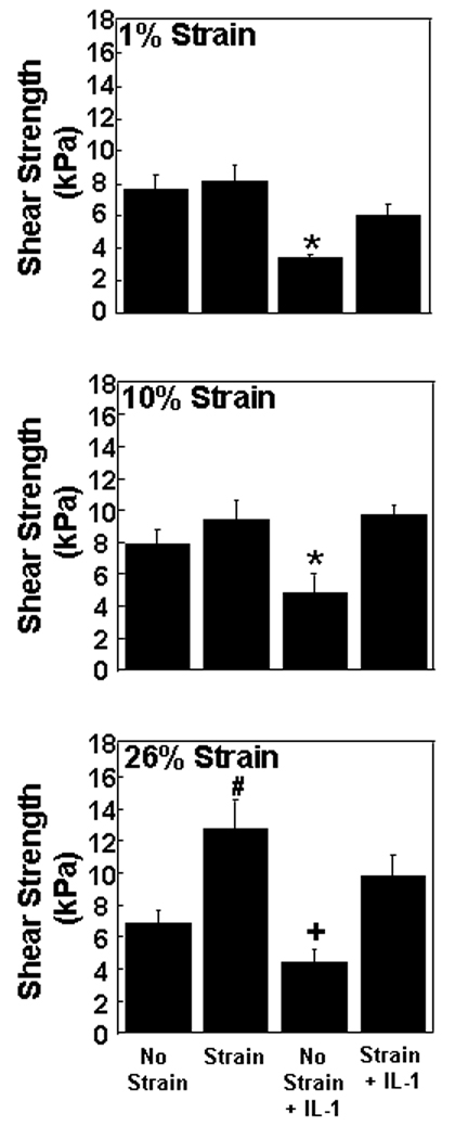
Results: IL-1 increased MMP activity, S-GAG release, and NO production, while decreasing the shear strength and tissue repair in the interface. Dynamic loading antagonized IL-1-mediated inhibition of repair at all strain amplitudes. Neither IL-1 treatment nor strain altered aggrecanase activity. Additionally, strain alone did not alter meniscal healing, except at the highest strain magnitude (26%), a level that enhanced the strength of repair.
Conclusions: Dynamic loading blocked the catabolic effects of IL-1 on meniscal repair, suggesting that joint loading through physical therapy may be beneficial in promoting healing of meniscal lesions under inflammatory conditions.
Copyright 2010 Osteoarthritis Research Society International. All rights reserved.
Conflict of interest statement
None of the authors have any financial or personal relationships with other people or organizations that influence this work.
Figures
Similar articles
-
Inhibition of integrative repair of the meniscus following acute exposure to interleukin-1 in vitro.J Orthop Res. 2008 Apr;26(4):504-12. doi: 10.1002/jor.20538. J Orthop Res. 2008. PMID: 18050309 Free PMC article.
-
Interleukin-1 and tumor necrosis factor alpha inhibit repair of the porcine meniscus in vitro.Osteoarthritis Cartilage. 2007 Sep;15(9):1053-60. doi: 10.1016/j.joca.2007.03.003. Epub 2007 Apr 19. Osteoarthritis Cartilage. 2007. PMID: 17448702 Free PMC article.
-
Enhanced integrative repair of the porcine meniscus in vitro by inhibition of interleukin-1 or tumor necrosis factor alpha.Arthritis Rheum. 2007 Sep;56(9):3033-42. doi: 10.1002/art.22839. Arthritis Rheum. 2007. PMID: 17729298
-
Meniscal repair using the outside-to-inside technique.Clin Sports Med. 1996 Jul;15(3):469-81. Clin Sports Med. 1996. PMID: 8800530 Review.
-
Treatment of meniscal injury: a current concept review.Chin J Traumatol. 2010 Dec;13(6):370-6. Chin J Traumatol. 2010. PMID: 21126396 Review.
Cited by
-
Molecular Biology of Meniscal Healing: A Narrative Review.Int J Mol Sci. 2024 Jan 7;25(2):768. doi: 10.3390/ijms25020768. Int J Mol Sci. 2024. PMID: 38255841 Free PMC article. Review.
-
Meniscus-Derived Matrix Scaffolds Promote the Integrative Repair of Meniscal Defects.Sci Rep. 2019 Jun 18;9(1):8719. doi: 10.1038/s41598-019-44855-3. Sci Rep. 2019. PMID: 31213610 Free PMC article.
-
Shedding light on the effects of blood on meniscus tissue: the role of mononuclear leukocytes in mediating meniscus catabolism.Osteoarthritis Cartilage. 2024 Aug;32(8):938-949. doi: 10.1016/j.joca.2024.04.022. Epub 2024 May 21. Osteoarthritis Cartilage. 2024. PMID: 38782253 Free PMC article.
-
TRPV4 differentially controls inflammatory cytokine networks during static and dynamic compression of the intervertebral disc.JOR Spine. 2023 Oct 27;6(4):e1282. doi: 10.1002/jsp2.1282. eCollection 2023 Dec. JOR Spine. 2023. PMID: 38156056 Free PMC article.
-
Reduced response of human meniscal cells to Osteogenic Protein 1 during osteoarthritis and pro-inflammatory stimulation.Osteoarthritis Cartilage. 2016 Jun;24(6):1036-46. doi: 10.1016/j.joca.2015.12.017. Epub 2016 Jan 8. Osteoarthritis Cartilage. 2016. PMID: 26778533 Free PMC article.
References
-
- Markolf KL, Bargar WL, Shoemaker SC, Amstutz HC. The role of joint load in knee stability. J Bone Joint Surg Am. 1981;63:570–585. - PubMed
-
- Ahmed AM, Burke DL. In-vitro measurement of static pressure distribution in synovial joints--Part I: Tibial surface of the knee. J Biomech Eng. 1983;105:216–225. - PubMed
-
- Wojtys EM, Chan DB. Meniscus Structure and Function. AAOS Instructional Course Lectures. 2005;54:323–330. - PubMed
-
- Spilker RL, Donzelli PS, Mow VC. A transversely isotropic biphasic finite element model of the meniscus. J Biomech. 1992;25:1027–1045. - PubMed
-
- Zielinska B, Donahue TL. 3D finite element model of meniscectomy: changes in joint contact behavior. J Biomech Eng. 2006;128:115–123. - PubMed
Publication types
MeSH terms
Substances
Grants and funding
LinkOut - more resources
Full Text Sources
