

doi: 10.1016/j.rdc.2009.12.003.
Cell-bound complement biomarkers for systemic lupus erythematosus: from benchtop to bedside
Affiliations
- PMID: 20202597
- PMCID: PMC2837510
- DOI: 10.1016/j.rdc.2009.12.003
Item in Clipboard
Cell-bound complement biomarkers for systemic lupus erythematosus: from benchtop to bedside
Rheum Dis Clin North Am.
2010 Feb.
Abstract
Systemic lupus erythematosus is arguably the most clinically and serologically diverse autoimmune disease. This article highlights the biomarkers helpful in diagnosing this disease. The authors' own research is presented.
Copyright 2010 Elsevier Inc. All rights reserved.
Figures
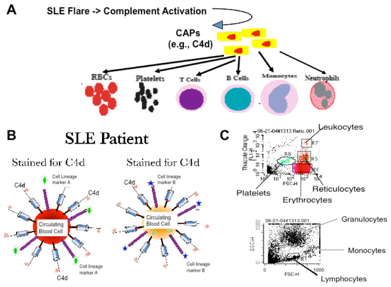
(A) Rationale: During SLE flares, considerable amounts of complement activation products may be generated. These CAPs may bind stably to various circulating cells in proportion to the extent of complement activation. (B) Schematic illustration of the multicolor staining of circulating cells for cell type-specific surface markers and surface-bound CAPs (e.g., C4d). (C) Representative dotplots demonstrate the identification of erythrocytes, reticulocytes, platelets, lymphocytes, monocytes, and granulocytes. These cell types can also be differentiated using cell lineage-specific mAbs added to the cell suspension (e.g., anti-CD3, anti-CD19, etc).
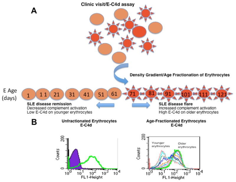
(A) Schematic illustration of the hypothetical model; (B) Histograms of E-C4d levels on age-fractionated erythrocytes prepared from a representative SLE patient. The open histograms represent the C4d staining on the entire population (left panel) or density/age-fractionated erythroctyes (right panel). The purple closed peak depicts the background staining of erythrocytes using an istotype mouse IgG control.
References
-
- Sherer Y, Gorstein A, Fritzler MJ, Shoenfeld Y. Autoantibody explosion in systemic lupus erythematosus: more than 100 different antibodies found in SLE patients. [see comment] Seminars in Arthritis & Rheumatism. 2004;34(2):501–37. - PubMed
-
- Rahman A, Isenberg DA. Systemic lupus erythematosus. N Engl J Med. 2008;358(9):929–39. - PubMed
-
- Tan EM, Cohen AS, Fries JF. The 1982 revised criteria for the classification of systemic lupus erythematosus. Arthritis & Rheumatism. 1982;25:1271–1277. - PubMed
-
- Hochberg MC. Updating the American College of Rheumatology revised criteria for the classification of systemic lupus erythematosus. Arthritis & Rheumatism. 1997;40:1725. - PubMed
-
- Bombardier C, Gladman DD, et al. Derivation of the SLEDAI. A disease activity index for lupus patients. The Committee on Prognosis Studies in SLE. Arthritis & Rheumatism. 1992;35:630–640. - PubMed
Publication types
MeSH terms
Substances
Grants and funding
- R01 AR046588/AR/NIAMS NIH HHS/United States
- R01AI077591/AI/NIAID NIH HHS/United States
- UL1 TR000005/TR/NCATS NIH HHS/United States
- K24 AR-02213/AR/NIAMS NIH HHS/United States
- K24 AR002213/AR/NIAMS NIH HHS/United States
- R01 AR-46588/AR/NIAMS NIH HHS/United States
- K23 AR-051044/AR/NIAMS NIH HHS/United States
- R01HL074335/HL/NHLBI NIH HHS/United States
- R01 HL074335/HL/NHLBI NIH HHS/United States
- K23 AR051044/AR/NIAMS NIH HHS/United States
- R01 AI077591/AI/NIAID NIH HHS/United States
- U01 AI077995/AI/NIAID NIH HHS/United States
LinkOut - more resources
Full Text Sources
Other Literature Sources
Medical
