Review
. 2010 Jul;35(7):411-8.
doi: 10.1016/j.tibs.2010.02.003. Epub 2010 Mar 2.

Structure, biological functions and applications of the AB5 toxins

Affiliations
Review

Structure, biological functions and applications of the AB5 toxins

Travis Beddoe et al. Trends Biochem Sci. 2010 Jul.

Abstract

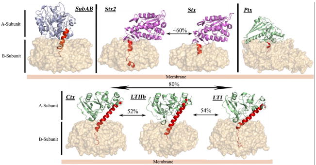
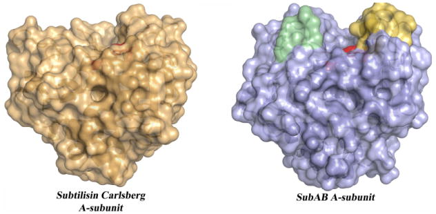
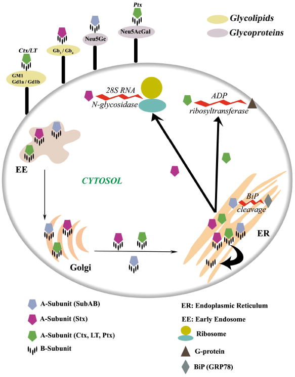
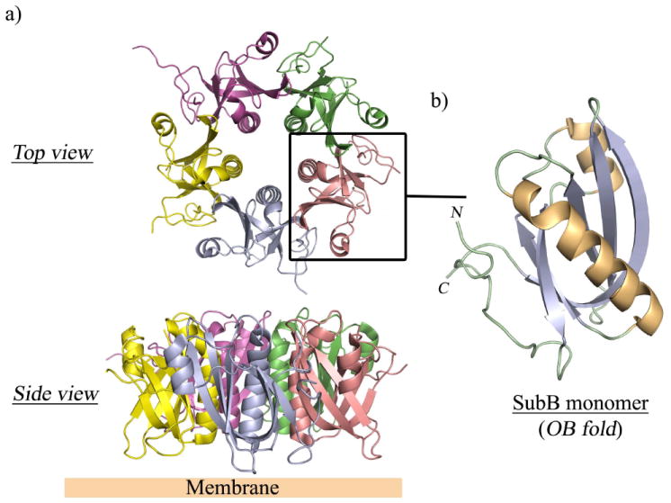
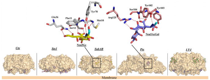
AB(5) toxins are important virulence factors for several major bacterial pathogens, including Bordetella pertussis, Vibrio cholerae, Shigella dysenteriae and at least two distinct pathotypes of Escherichia coli. The AB(5) toxins are so named because they comprise a catalytic A-subunit, which is responsible for disruption of essential host functions, and a pentameric B-subunit that binds to specific glycan receptors on the target cell surface. The molecular mechanisms by which the AB(5) toxins cause disease have been largely unravelled, including recent insights into a novel AB(5) toxin family, subtilase cytotoxin (SubAB). Furthermore, AB(5) toxins have become a valuable tool for studying fundamental cellular functions, and are now being investigated for potential applications in the clinical treatment of human diseases.


Figures

Figure 1
Figure 1. Crystal structures of members of the four recognised AB5 toxin families
The B-subunit is represented as molecular surface. The A-subunits of SubAB, Ctx and LT, Stx and Ptx are shown in cartoon representation and coloured according to the respective catalytic activity (Light blue for subtilase activity, light green for ADP-ribosylase activity and purple for RNA N-glycosidase activity). The common structural element (Helix A2) is coloured in red, and the level of sequence identity of the A-subunit inside a family is also indicated.
Figure 2
Figure 2. Molecular surface representations of a broad specificity protease and the SubAB A-subunit
The position of the catalytic triad (His-Asp-Ser) in both X-ray structures is coloured in red. The Subtilisin Carlsberg (Pdb code: 1C3L) is a typical broad specificity protease with a shallow active site. The loops composed of the amino acid residues 81-88 and 234-239 forming a deeper binding cleft in SubAB are coloured in yellow and pale green, respectively.
Figure 3
Figure 3. [SC1]Schematic representation of the glycan surface recognition of the various AB5 toxins and their respective trafficking pathways into the cell
The toxins’ A-subunits are shown as a pentagon and each subunit is coloured according to the specific catalytic activity (blue for protease activity, green and magenta for ADP-ribosyltransferase and N-glycosidase activities, respectively). The toxins bind to the cell-surface via their respective glycan receptors, where they are internalized to the early endosomal (EE) compartment. The names of these glycan receptors are indicated and inserted into schematic rings located on the surface of the cell and coloured according to their nature (grey and pale green for glycoproteins and glycolipids, respectively). The toxins are then trafficked to the Golgi then onto the endoplasmic reticulum (ER) where the A-subunit of the Stx, Ctx, LT and Ptx are separated from the B-pentamer. They are then exported out of the ER, where they are able to attack their respective molecular targets (the G-protein (brown triangle), the 28S ribosomal RNA, and the ribosome (teal[SC2]). In the case of SubAB, it is unknown whether SubA is separated from SubB; however the purified SubA and SubAB can cleave BiP (grey) in vitro .
Figure 4
Figure 4. The pentameric B-subunit of the AB5 toxin
a) Overall architecture of the pentameric B-subunit of SubAB shown in two different orientations. Each monomer is coloured differently. b) Close up view of a monomer forming the common oligosaccharide/oligonucleotide (OB) fold. The monomer is coloured according to the secondary structural elements (helices, gold; β-strands, purple).
Figure 5
Figure 5. Molecular surface representations of the B-subunits of Ctx, Stx1, SubAB, Ptx and LT-I with bound glycans
The Gb3 analogue (PK-MCO) is shown bound to Stx1. The antagonist BMSC-0011 (in blue) is shown bound to LT-I and Ctx. The second binding site of LT-I is also shown with the blood group A antigen analogue (in green) bound in a similar position as in SubAB, whereas Neu5Gc and Neu5Acα-3Gal are shown bound to SubAB and Ptx, respectively. A close-up view of the interactions for SubAB–Neu5Gc and Ptx–Neu5Acα2-3Gal is also represented. For clarity, only the amino acid residues involved in direct hydrogen bonds (dashed red lines) with the two ligands are shown. The position of the extra hydroxyl in Neu5Gc and the equivalent position in Neu5Acα2-3Gal are highlighted in cyan.

Similar articles

Cited by

References

    1. Sack DA, et al. Cholera. Lancet. 2004;363:223–233. - PubMed
    1. Nataro JP, Kaper JB. Diarrheagenic Escherichia coli. Clin Microbiol Rev. 1998;11:142–201. - PMC - PubMed
    1. Paton JC, Paton AW. Pathogenesis and Diagnosis of Shiga Toxin-Producing Escherichia coli Infections. Clinical Microbiology Reviews. 1998;11:450–479. - PMC - PubMed
    1. Tarr PI, et al. Shiga-toxin-producing Escherichia coli and haemolytic uraemic syndrome. Lancet. 2005;365:1073–1086. - PubMed
    1. Griffin P. Shiga toxin-producing Escherichia coli infections in the United States. 4th International Symposium and Workshop on Shiga Toxin-producing Escherichia coli Infections. 2000:21.

Publication types

LinkOut - more resources