

Reversal of impaired myocardial beta-adrenergic receptor signaling by continuous-flow left ventricular assist device support
- PMID: 20202864
- PMCID: PMC2876229
- DOI: 10.1016/j.healun.2010.01.010
Reversal of impaired myocardial beta-adrenergic receptor signaling by continuous-flow left ventricular assist device support
Abstract
Background: Myocardial beta-adrenergic receptor (beta-AR) signaling is severely impaired in chronic heart failure (HF). This study was conducted to determine if left ventricular (LV) beta-AR signaling could be restored after continuous-flow LV assist device (LVAD) support.
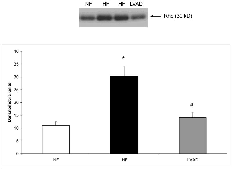
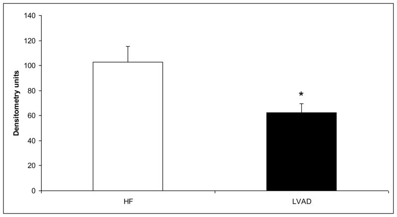
Methods: Twelve patients received LVADs as a bridge to transplant. Paired LV biopsy specimens were obtained at the time of LVAD implant (HF group) and transplant (LVAD group). The mean duration of LVAD support was 152 +/- 34 days. Myocardial beta-AR signaling was assessed by measuring adenylyl cyclase (AC) activity, total beta-AR density (B(max)), and G protein-coupled receptor kinase-2 (GRK2) expression and activity. LV specimens from 8 non-failing hearts (NF) were used as controls.
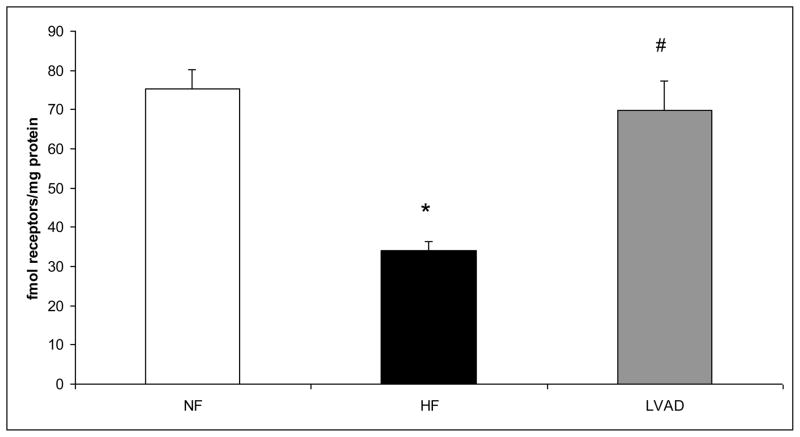
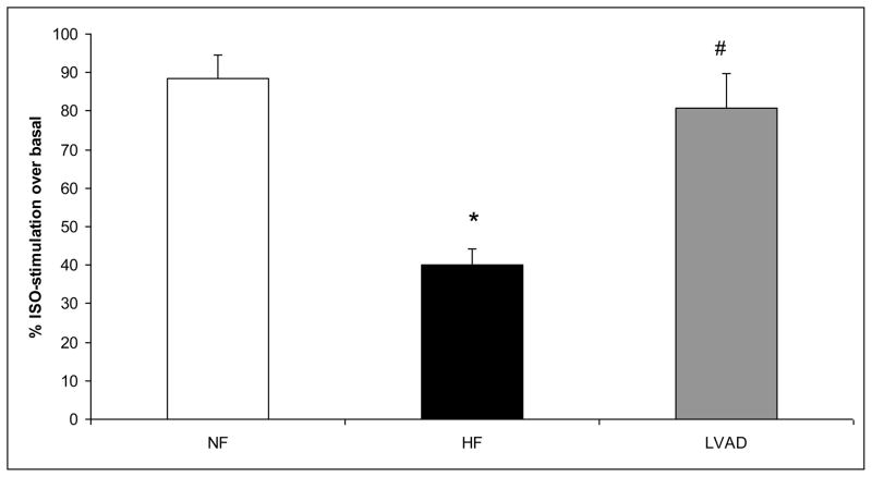
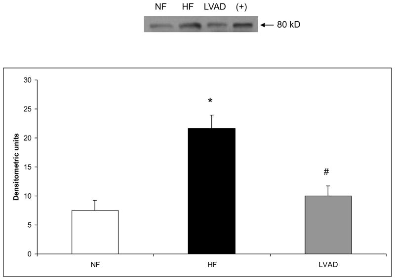
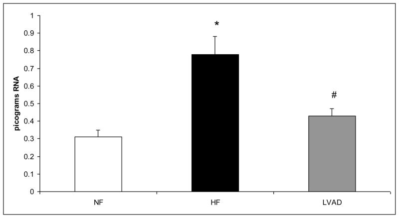
Results: Basal and isoproterenol-stimulated AC activity was significantly lower in HF vs NF, indicative of beta-AR uncoupling. Continuous-flow LVAD support restored basal and isoproterenol-stimulated AC activity to levels similar to NF. B(max) was decreased in HF vs NF and increased to nearly normal in the LVAD group. GRK2 expression was increased 2.6-fold in HF vs NF and was similar to NF after LVAD support. GRK2 activity was 3.2-fold greater in HF vs NF and decreased to NF levels in the LVAD group.
Conclusions: Myocardial beta-AR signaling can be restored to nearly normal after continuous-flow LVAD support. This is similar to previous data for volume-displacement pulsatile LVADs. Decreased GRK2 activity is an important mechanism and indicates that normalization of the neurohormonal milieu associated with HF is similar between continuous-flow and pulsatile LVADs. This may have important implications for myocardial recovery.
Copyright 2010 International Society for Heart and Lung Transplantation. Published by Elsevier Inc. All rights reserved.
Figures
Similar articles
-
Lymphocyte levels of GRK2 (betaARK1) mirror changes in the LVAD-supported failing human heart: lower GRK2 associated with improved beta-adrenergic signaling after mechanical unloading.J Card Fail. 2006 Jun;12(5):360-8. doi: 10.1016/j.cardfail.2006.02.011. J Card Fail. 2006. PMID: 16762799
-
Restoration of myocardial beta-adrenergic receptor signaling after left ventricular assist device support.J Thorac Cardiovasc Surg. 2006 May;131(5):975-80. doi: 10.1016/j.jtcvs.2006.01.027. J Thorac Cardiovasc Surg. 2006. PMID: 16678578
-
Molecular Changes in Children with Heart Failure Undergoing Left Ventricular Assist Device Therapy.J Pediatr. 2017 Mar;182:184-189.e1. doi: 10.1016/j.jpeds.2016.11.011. Epub 2016 Nov 29. J Pediatr. 2017. PMID: 27908653 Free PMC article.
-
GRK2 in the heart: a GPCR kinase and beyond.Antioxid Redox Signal. 2014 Nov 10;21(14):2032-43. doi: 10.1089/ars.2014.5876. Epub 2014 May 13. Antioxid Redox Signal. 2014. PMID: 24702056 Free PMC article. Review.
-
Reverse remodeling following insertion of left ventricular assist devices (LVAD): a review of the morphological and molecular changes.Cardiovasc Res. 2005 Dec 1;68(3):376-86. doi: 10.1016/j.cardiores.2005.06.030. Epub 2005 Jul 18. Cardiovasc Res. 2005. PMID: 16024006 Review.
Cited by
-
Novel Molecular Approaches in Heart Failure: Seven Trans-Membrane Receptors Signaling in the Heart and Circulating Blood Leukocytes.Front Cardiovasc Med. 2015 Mar 16;2:13. doi: 10.3389/fcvm.2015.00013. eCollection 2015. Front Cardiovasc Med. 2015. PMID: 26664885 Free PMC article. Review.
-
Reverse Remodeling With Left Ventricular Assist Devices.Circ Res. 2021 May 14;128(10):1594-1612. doi: 10.1161/CIRCRESAHA.121.318160. Epub 2021 May 13. Circ Res. 2021. PMID: 33983828 Free PMC article. Review.
-
Myocardial recovery evaluation from ventricular assist device in patients with dilated cardiomyopathy.ESC Heart Fail. 2022 Aug;9(4):2491-2499. doi: 10.1002/ehf2.13951. Epub 2022 May 10. ESC Heart Fail. 2022. PMID: 35535672 Free PMC article.
-
Left ventricular reverse remodelling and its predictors in non-ischaemic cardiomyopathy.ESC Heart Fail. 2022 Aug;9(4):2070-2083. doi: 10.1002/ehf2.13939. Epub 2022 Apr 18. ESC Heart Fail. 2022. PMID: 35437948 Free PMC article. Review.
-
G protein coupled receptor kinases as therapeutic targets in cardiovascular disease.Circ Res. 2011 Jul 22;109(3):309-19. doi: 10.1161/CIRCRESAHA.110.231233. Circ Res. 2011. PMID: 21778435 Free PMC article. Review.
References
-
- Bristow MR, Ginsburg R, Minobe W, et al. Decreased catecholamine sensitivity and beta-adrenergic-receptor density in failing human hearts. N Engl J Med. 1982;307:205–211. - PubMed
-
- Brodde OE, Michel MC, Zerkowski HR. Signal transduction mechanisms controlling cardiac contractility and their alterations in chronic heart failure. Cardiovasc Res. 1995;30:570–584. - PubMed
-
- Inglese J, Freedman NJ, Koch WJ, et al. Structure and mechanism of the G protein-coupled receptor kinases. J Biol Chem. 1993;268:23735–23738. - PubMed
-
- Ungerer M, Parruti G, Bohm M, et al. Expression of beta-arrestins and beta-adrenergic receptor kinases in the failing human heart. Circ Res. 1994;74:206–213. - PubMed
-
- Ungerer M, Bohm M, Elce JS, et al. Altered expression of beta-adrenergic receptor kinase and beta 1-adrenergic receptors in the failing human heart. Circulation. 1993;87:454–463. - PubMed
Publication types
MeSH terms
Substances
Grants and funding
LinkOut - more resources
Full Text Sources
Medical
Research Materials
Miscellaneous
