. 2010 Apr 30;285(18):13788-96.
doi: 10.1074/jbc.M109.066670. Epub 2010 Mar 4.

Mapping of domains on HIV envelope protein mediating association with calnexin and protein-disulfide isomerase

Affiliations

Mapping of domains on HIV envelope protein mediating association with calnexin and protein-disulfide isomerase

Marie-Jeanne Papandréou et al. J Biol Chem. .

Abstract

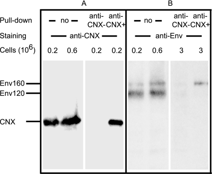
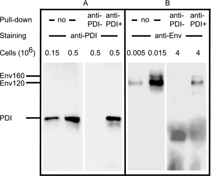
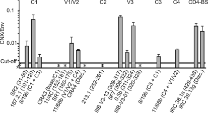
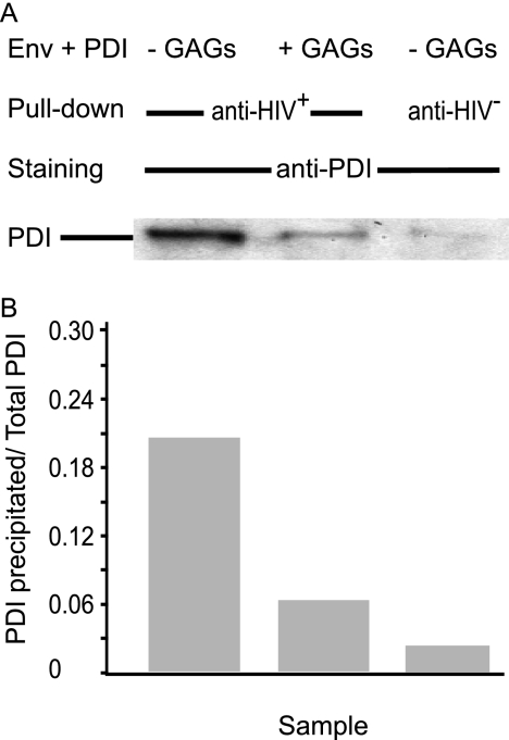
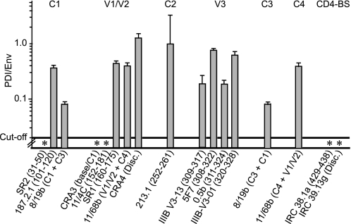
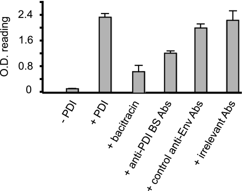
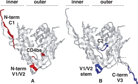
The cell catalysts calnexin (CNX) and protein-disulfide isomerase (PDI) cooperate in establishing the disulfide bonding of the HIV envelope (Env) glycoprotein. Following HIV binding to lymphocytes, cell-surface PDI also reduces Env to induce the fusogenic conformation. We sought to define the contact points between Env and these catalysts to illustrate their potential as therapeutic targets. In lysates of Env-expressing cells, 15% of the gp160 precursor, but not gp120, coprecipitated with CNX, whereas only 0.25% of gp160 and gp120 coprecipitated with PDI. Under in vitro conditions, which mimic the Env/PDI interaction during virus/cell contact, PDI readily associated with Env. The domains of Env interacting in cellulo with CNX or in vitro with PDI were then determined using anti-Env antibodies whose binding site was occluded by CNX or PDI. Antibodies against domains V1/V2, C2, and the C terminus of V3 did not bind CNX-associated Env, whereas those against C1, V1/V2, and the CD4-binding domain did not react with PDI-associated Env. In addition, a mixture of the latter antibodies interfered with PDI-mediated Env reduction. Thus, Env interacts with intracellular CNX and extracellular PDI via discrete, largely nonoverlapping, regions. The sites of interaction explain the mode of action of compounds that target these two catalysts and may enable the design of further new competitive agents.


Figures

FIGURE 1.
FIGURE 1.
Coprecipitation of CNX and PDI with Env using anti-Env Abs. Lysate of BHK-21 cells (the corresponding cell number is indicated) expressing Env was submitted (no pulldown) to SDS-PAGE analysis and WB using anti-Env (A), -CNX (B), or -PDI (C) Abs. Alternatively, lysate was incubated with irrelevant sheep IgG (anti-Env) or D7324 Ab (anti-Env+) prior to precipitation, elution using loading buffer, SDS-PAGE, and WB as indicated. A, *, nonspecific species detected by the peroxidase-labeled anti-sheep Ab staining system.
FIGURE 2.
FIGURE 2.
Coprecipitation of Env with CNX using anti-CNX Ab. Lysate of BHK-21 cells expressing Env was submitted (no pulldown) to SDS-PAGE analysis and WB using anti-CNX (A) or -Env (B) Abs. Alternatively, lysate was incubated with irrelevant rabbit IgG (anti-CNX) or anti-CNX Ab (anti-CNX+) prior to precipitation, elution using loading buffer, SDS-PAGE, and WB as indicated.
FIGURE 3.
FIGURE 3.
Coprecipitation of Env with PDI using anti-PDI Ab. Lysate of BHK-21 cells expressing Env was submitted (no pulldown) to SDS-PAGE analysis and WB using anti-PDI (A) or -Env (B) Abs. Alternatively, lysate was incubated with irrelevant mouse IgGs (anti-PDI) or anti-PDI Ab (anti-PDI+) prior to precipitation, elution using loading buffer and analysis by SDS-PAGE and WB as indicated.
FIGURE 4.
FIGURE 4.
CNX association with Env in cellulo, epitopes involved. Clarified lysate of BHK-21 cells expressing Env was incubated with dilutions of dot-blotted anti-Env Abs. The amount of Env associated with each dot was assessed using D7324 Ab or irrelevant sheep IgG prior to staining and densitometry. The presence of CNX associated with samples bound to the Abs was assessed in parallel using anti-CNX Ab or irrelevant rabbit IgGs. Env and CNX quantification was done using standard curves obtained using known amounts of antigens. For each Ab dilution, the CNX/Env molar ratio was determined (n = 2 experiments). The mean ratio ± S.D. obtained for each Ab is presented. * indicates Abs that did not bind Env complexed with CNX.
FIGURE 5.
FIGURE 5.
Glycosaminoglycans and PDI association with Env in vitro. Env was incubated with equimolar amounts of PDI in the presence or absence of glycosaminoglycans (GAGs) prior to precipitation using human HIV+ or HIV sera. The resulting samples were analyzed by SDS-PAGE and WB using anti-PDI Abs (A) prior to densitometric quantification (B) using a standard curve obtained using known amounts of PDI. The ratio “PDI recovered following precipitation/PDI in the assay” is shown.
FIGURE 6.
FIGURE 6.
PDI association with Env in vitro, epitopes involved. Env preincubated with PDI was incubated with dilutions of dot-blotted anti-Env Abs. The amount of Env associated with each dot was assessed using HIV+ or HIV sera prior to staining and densitometry. The presence of PDI associated with samples bound to the Abs was assessed in parallel using anti-PDI Ab, similar experiments using Env but not PDI determining the background signal. Env and PDI quantification was done using standard curves obtained using known amounts of antigens. For each Ab dilution, the PDI/Env molar ratio was determined (n = two experiments). The mean ratio ± S.D. obtained for each Ab is presented. * indicates Abs that did not bind Env complexed with PDI.
FIGURE 7.
FIGURE 7.
Effect of anti-Env Abs on reduction by PDI. Env coupled to Sepharose beads was incubated in the presence or absence of PDI. The sulfhydryl groups were then labeled using MPB, a thiol-reactive reagent that was detected using streptavidin-peroxidase and o-phenylenediamine. Bacitracin was used to inhibit Env reduction by PDI, as indicated. Alternatively, Env was incubated with anti-PDI-binding site Abs (a mixture of SR2, CRA3, 11/4c, IRC 38.1a, and IRC 39.13g), other anti-Env Abs (a mixture of 8/19b, SR1, 11.68, 213.1, and 5F7), or irrelevant mouse Abs, as described under “Experimental Procedures,” prior to reduction by PDI, MPB labeling, and processing (n = 3; the mean ratio ± S.D. is presented).
FIGURE 8.
FIGURE 8.
Location of the interaction sites for PDI and CNX in the mature gp120 structure. The gp120 structure shown was rendered from the recently released structure (Protein Data Bank code 3JWO (50)) that includes an extended N terminus when compared with earlier structures. The V3 loop in B was rendered from the single structure reporting it (Protein Data Bank code 2B4C) and then grafted where shown. The rendering is in the canonical view of gp120 (45) with the inner and outer domains of the molecule indicated. The definitive epitopes for the Abs that blocked chaperone binding are indicated, where available. PDI-reactive sites are largely accessible on the mature molecule (A), and those reacting with CNX are buried or face inward (B), consistent with access only prior to folding. The sites are mutually exclusive except for the V1/V2 region where precise residues cannot be indicated as they are not present in any solved structure. The diagram is illustrative, and the actual structure of the molecule during interaction with either chaperone remaining unknown.

Similar articles

Cited by

References

    1. Nagradova N. (2007) Curr. Protein Pept. Sci. 8, 273–282 - PubMed
    1. Dettenhofer M., Yu X. F. (2001) J. Biol. Chem. 276, 5985–5991 - PubMed
    1. Otteken A., Moss B. (1996) J. Biol. Chem. 271, 97–103 - PubMed
    1. Caramelo J. J., Parodi A. J. (2008) J. Biol. Chem. 283, 10221–10225 - PMC - PubMed
    1. Ihara Y., Cohen-Doyle M. F., Saito Y., Williams D. B. (1999) Mol. Cell 4, 331–341 - PubMed

Publication types

MeSH terms