. 2010 Apr 30;285(18):13951-7.
doi: 10.1074/jbc.M109.083378. Epub 2010 Mar 4.

On the relevance of the Met-turn methionine in metzincins

Affiliations

On the relevance of the Met-turn methionine in metzincins

Cynthia Tallant et al. J Biol Chem. .

Abstract

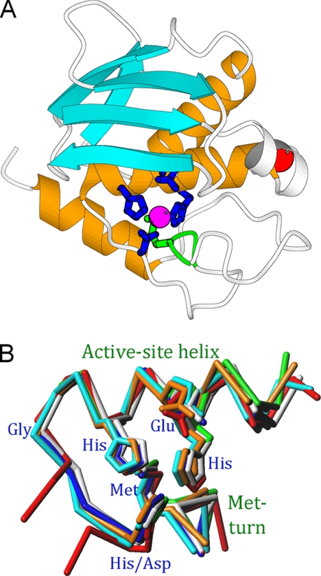
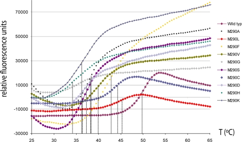
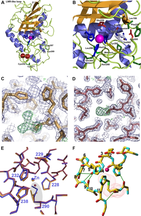
The metzincins are a clan of metallopeptidases consisting of families that share a series of structural elements. Among them is the Met-turn, a tight 1,4-turn found directly below the zinc-binding site, which is structurally and spatially conserved and invariantly shows a methionine at position 3 in all metzincins identified. The reason for this conservation has been a matter of debate since its discovery. We have studied this structural element in Methanosarcina acetivorans ulilysin, the structural prototype of the pappalysin family, by generating 10 mutants that replaced methionine with proteogenic amino acids. We compared recombinant overexpression yields, autolytic and tryptic activation, proteolytic activity, thermal stability, and three-dimensional structure with those of the wild type. All forms were soluble and could be purified, although with varying yields, and three variants underwent autolysis, could be activated by trypsin, and displayed significant proteolytic activity. All variants were analyzed for the thermal stability of their zymogens. None of the mutants analyzed proved more stable or active than the wild type. Both bulky and small side chains, as well as hydrophilic ones, showed diminished thermal stability. Two mutants, leucine and cysteine, crystallized and showed three-dimensional structures that were indistinguishable from the wild type. These studies reveal that the Met-turn acts as a plug that snugly inserts laterally into a core structure created by the protein segment engaged in zinc binding and thus contributes to its structural integrity, which is indispensable for function. Replacement of the methionine with residues that deviate in size, side-chain conformation, and chemical properties impairs the plug-core interaction and prejudices molecular stability and activity.


Figures

FIGURE 1.
FIGURE 1.
Common structural elements in metzincins. A, Richardson plot depicting the smallest metzincin catalytic domain structurally characterized, snapalysin from S. caespitosus (PDB 1C7K) (33), in standard orientation. The repetitive secondary structure elements common to all metzincins are shown in orange (helices) and cyan (β-strands), and the Met-turn is displayed in green. B, overlay of the seven metzincin prototypes structurally characterized to date as Cα plots showing the active-site helix (including the side chains of the zinc-liganding histidines and aspartate, and the general-base/acid glutamate) and the Met-turn (with the methionine side chain). The catalytic zinc has been omitted for clarity. Color coding: astacin, blue; adamalysin II, cyan; leishmanolysin, red; MMP-8, green; aeruginolysin, white; snapalysin, yellow; and ulilysin, orange (see 3, 5).
FIGURE 2.
FIGURE 2.
Thermal shift assays. Unfolding transition curves showing temperature-dependent change in fluorescence of the M290X variants analyzed. The Tm was obtained from the inflection point of each curve (see supplemental Table 3).
FIGURE 3.
FIGURE 3.
Ulilysin structure. A, Richardson-type plot of ulilysin M290L shown in standard orientation, i.e. with the view into the active-site cleft running left to right. Repetitive secondary structure elements are shown as violet ribbons for helices (labeled α1–α5) and as orange arrows for β-strands (labeled β1–β8). The catalytic zinc ion and the structural calcium ions are shown as magenta and red spheres, respectively. Selected residues engaged in zinc binding or catalysis and a dipeptide bound at the active-site cleft (Arg501-Val502) are shown as stick models. B, close-up view of A around the Met-turn. C, detail of the initial σA-weighted (2mFo − DFc)-type (violet mesh; contoured at 1σ) and (mFo − DFc)-type (green mesh; contoured at 3σ) omit electron-density maps of ulilysin M290L centered on the latter residue. The initial model used for phasing (with an alanine at this position) is superimposed for orientation. D, same as C but for ulilysin M290C. E, superposition of the polypeptide chain traces of ulilysin C269A (blue), M290L (orange), and M290C (mauve). Position 290 is highlighted by a gray ellipse. F, structure of the conserved metzincin core that maintains structural integrity of the zinc-binding site. Hydrogen bonds are shown as green dashed lines, the positions within the zinc-binding consensus sequence (1–11) are labeled, and the van-der-Waals surface of the Met-turn methionine is depicted with red dots.

Similar articles

Cited by

References

    1. Scheef E. D., Fink J. L. (2003) in Structural Bioinformatics (Bourne P. E., Weissig H. eds) pp. 15–39, Wiley-Liss, Inc., Hoboken, NJ
    1. Bode W., Gomis-Rüth F. X., Stöckler W. (1993) FEBS Lett. 331, 134–140 - PubMed
    1. Gomis-Rüth F. X. (2003) Mol. Biotech. 24, 157–202 - PubMed
    1. Stöcker W., Grams F., Baumann U., Reinemer P., Gomis-Rüth F. X., McKay D. B., Bode W. (1995) Protein Sci. 4, 823–840 - PMC - PubMed
    1. Gomis-Rüth F. X. (2009) J. Biol. Chem. 284, 15353–15357 - PMC - PubMed

Publication types

MeSH terms

LinkOut - more resources