. 2010 Apr 30;285(18):13380-7.
doi: 10.1074/jbc.M109.061366. Epub 2010 Mar 3.

Functional oligomerization of the Saccharomyces cerevisiae isoprenylcysteine carboxyl methyltransferase, Ste14p

Affiliations

Functional oligomerization of the Saccharomyces cerevisiae isoprenylcysteine carboxyl methyltransferase, Ste14p

Amy M Griggs et al. J Biol Chem. .

Abstract

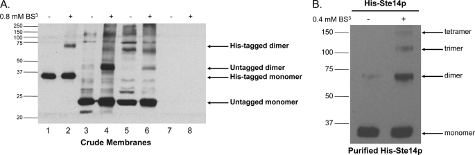
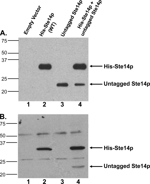
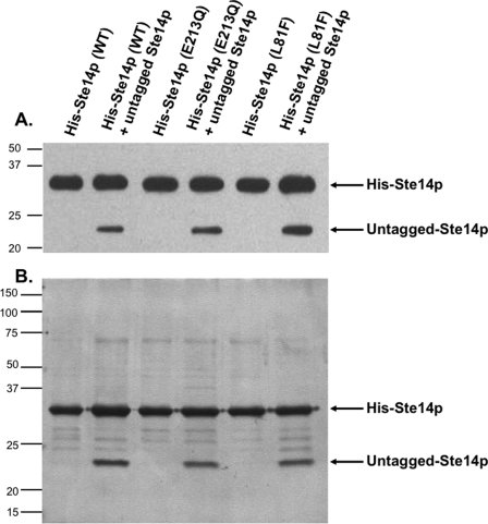
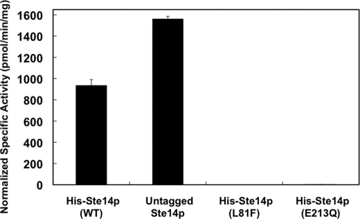
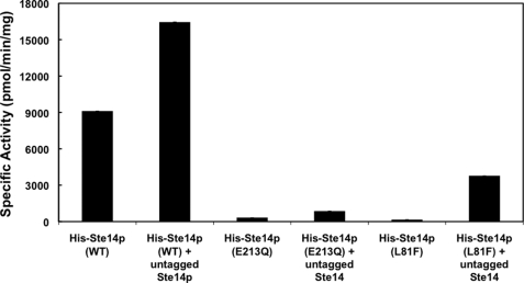
The isoprenylcysteine carboxyl methyltransferase (Icmt) from Saccharomyces cerevisiae, also designated Ste14p, is a 26-kDa integral membrane protein that contains six transmembrane spanning segments. This protein is localized to the endoplasmic reticulum membrane where it performs the methylation step of the CAAX post-translational processing pathway. Sequence analysis reveals a putative GXXXG dimerization motif located in transmembrane 1 of Ste14p, but it is not known whether Ste14p forms or functions as a dimer or higher order oligomer. We determined that Ste14p predominantly formed a homodimer in the presence of the cross-linking agent, bis-sulfosuccinimidyl suberate. Wild-type untagged Ste14p also co-immunoprecipitated and co-purified with N-terminal-tagged His(10)-myc(3)-Ste14p (His-Ste14p). Furthermore, enzymatically inactive His-Ste14p variants L81F and E213Q both exerted a dominant-negative effect on methyltransferase activity when co-expressed and co-purified with untagged wild-type Ste14p. Together, these data, although indirect, suggest that Ste14p forms and functions as a homodimer or perhaps a higher oligomeric species.


Figures

FIGURE 1.
FIGURE 1.
Immunoblot of His-Ste14p cross-linked chemically with BS3. A, 80 μg of crude membrane protein were incubated with 0.8 mm BS3 for 20 min at room temperature. The reaction was terminated by the addition of SDS-PAGE sample buffer without reducing agents and then heated for 20 min at 65 °C. Various amounts of crude membrane proteins were separated by 10% SDS-PAGE, and His-Ste14p was detected using a Ste14p polyclonal antibody (1:1,000) and a HRP-conjugated secondary goat anti-rabbit antibody (1:10,000) as follows: lane 1, 1 μg of His-Ste14p (CH2704); lane 2, 1 μg of His-Ste14p (CH2704) + BS3; lane 3, 10 μg of untagged Ste14p (CH2866; 2μ PGK promoter); lane 4, 10 μg of untagged Ste14p (CH2866; 2μ PGK promoter) + BS3; lane 5, 50 μg of SM3495 (untagged Ste14p; 2μ STE14 promoter); lane 6, 50 μg of SM3495 (untagged Ste14p; 2μ STE14 promoter) + BS3; lane 7, 50 μg of CH2714 (negative control); lane 8, 50 μg of CH2714 (negative control) + BS3. Protein bands were visualized using enhanced chemiluminescence (ECL). B, purified His-Ste14p (2.5 μg) was incubated with 0.4 mm BS3 for 20 min, and the reaction was terminated by the addition of SDS-PAGE sample buffer without reducing agents. The reactions were heated for 30 min at 65 °C, 0.1 μg of purified protein was separated by 7.5% SDS-PAGE, and the protein bands were visualized by immunoblot analysis as described above.
FIGURE 2.
FIGURE 2.
Expression and co-immunoprecipitation of untagged Ste14p with His-Ste14p. A, 2 μg of crude membrane protein expressing tagged or untagged Ste14p variants were subjected to 10% SDS-PAGE and immunoblot analyses. First lane, empty vector; second lane, His-Ste14p; third lane, untagged-Ste14p; fourth lane, His-Ste14p + untagged-Ste14p. Proteins were detected with a Ste14p polyclonal antibody (1:500) and HRP-conjugated goat anti-rabbit secondary antibody (1:10,000), and the bands were visualized using ECL. B, 100 μg of protein from crude membrane preparations were incubated in 1× RIPA buffer plus ∼3 μg of anti-Myc monoclonal antibody overnight at 4 °C with gentle rotation. 40 μl of 50% (vol:vol) protein A-Sepharose beads in 1× RIPA buffer were added to each tube and incubated another 2.5 h at 4 °C with gentle rotation. The bead-immuno-protein complexes were processed as described under “Experimental Procedures,” and the entire sample was separated by 10% SDS-PAGE. Immunodetection was with a Ste14p polyclonal antibody (1:500) and a goat anti-rabbit secondary antibody (1:10,000) conjugated to HRP. Protein bands were visualized by ECL. WT, wild type.
FIGURE 3.
FIGURE 3.
Co-expression and co-purification of untagged Ste14p with His-Ste14p and inactive His-Ste14p variants L81F and E213Q. A, expression analysis is shown. 2 μg of crude membrane protein expressing tagged or untagged Ste14p variants were subjected to 10% SDS-PAGE and immunoblot analyses. Proteins were detected with a Ste14p polyclonal antibody (1:500) and HRP-conjugated goat anti-rabbit secondary antibody (1:10,000), and the bands were visualized using ECL. B, co-purification is shown. 25 μg of crude membrane protein was solubilized in 1% (w/v) DDM for 1 h and centrifuged at 300,000 × g for 30 min at 4 °C to remove un-solubilized protein. The supernatant was incubated with Talon metal affinity resin, and the resin was washed as described under “Experimental Procedures.” The purified proteins were eluted with 1 m imidazole, and the fractions were combined and concentrated. 1 μg of purified protein was subjected to 10% SDS-PAGE analysis, and the proteins were visualized by silver nitrate staining. WT, wild type.
FIGURE 4.
FIGURE 4.
In vitro methyltransferase activities of Ste14p variants in crude membranes. 5 μg of crude membrane protein were incubated with 200 μm AFC and 20 μm [14C]SAM for 30 min at 30 °C in a total volume of 60 μl. The reactions were terminated by the addition of 50 μl of 1 m NaOH/1% SDS (v/v), and the activity was quantified by the vapor diffusion assay as described under “Experimental Procedures.” Each reaction was performed three times in duplicate, and error bars represent ±S.D.). Activities were normalized to the expression level of each construct using immunoblots containing varying amounts of each protein. The protein bands were visualized with ECL as described in Fig. 3 and quantified using a Nucleovision imaging system equipped with a high sensitivity cooled CCD camera. WT, wild type.
FIGURE 5.
FIGURE 5.
In vitro methyltransferase activities of purified His-Ste14p and variants. Purified protein (0.3 μg) was reconstituted in a 3000-fold excess of E. coli polar lipid by 20-fold rapid dilution in 100 mm MES, pH 7.0, and then incubated on ice for 10 min. Reactions contained the reconstituted purified proteins, 200 μm AFC, and 20 μm [14C]SAM and were incubated for 30 min at 30 °C. The reactions were terminated by the addition of 1 m NaOH, 1% SDS, and activity was quantified as described under “Experimental Procedures.” Each reaction was performed three times in duplicate, and error bars represent ±S.D. WT, wild type.

Similar articles

Cited by

References

    1. Clarke S. (1992) Annu. Rev. Biochem. 61, 355–386 - PubMed
    1. Zhang F. L., Casey P. J. (1996) Annu. Rev. Biochem. 65, 241–269 - PubMed
    1. Young S. G., Ambroziak P., Kim E., Clarke S. (2000) The Enzymes: Protein Lipidation (Tamanoi F., Sigman D. S. eds) Vol. 21, pp. 155–213, Academic Press, San Diego, CA
    1. Hrycyna C. A., Clarke S. (1993) Pharmacol. Ther. 59, 281–300 - PubMed
    1. Gelb M. H., Brunsveld L., Hrycyna C. A., Michaelis S., Tamanoi F., Van Voorhis W. C., Waldmann H. (2006) Nat. Chem. Biol. 2, 518–528 - PMC - PubMed

MeSH terms

LinkOut - more resources