

Caveolae, caveolins, and cavins: complex control of cellular signalling and inflammation
- PMID: 20202978
- PMCID: PMC2856194
- DOI: 10.1093/cvr/cvq075
Caveolae, caveolins, and cavins: complex control of cellular signalling and inflammation
Abstract
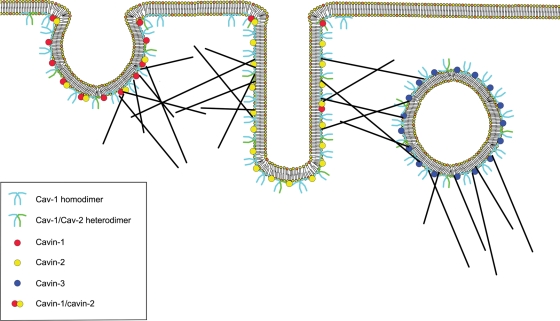
Caveolae are specialized lipid rafts that form flask-shaped invaginations of the plasma membrane. They are involved in cell signalling and transport and have been shown critically regulate vascular reactivity and blood pressure. The organization and functions of caveolae are mediated by coat proteins (caveolins) and support or adapter proteins (cavins). The caveolins, caveolin-1, -2, and -3, form the structural backbone of caveolae. These proteins are also highly integrated into caveolae function and have their own activity independent of caveolae. The cavins, cavins 1-4, are involved in regulation of caveolae and modulate the function of caveolins by promoting the membrane remodelling and trafficking of caveolin-derived structures. The relationships between these different proteins are complex and intersect with many aspects of cell function. Caveolae have also been implicated in chronic inflammatory conditions and other pathologies including atherosclerosis, inflammatory bowel disease, muscular dystrophy, and generalized dyslipidaemia. The pathogenic role of the caveolins is an emerging area, however, the roles of cavins in disease is just beginning to be explored. This review will examine the relationship between caveolins and cavins and explore the role of caveolae in inflammatory signalling mechanisms.
Figures
References
-
- Palade GE. Fine structure of blood capillaries. J Appl Phys. 1953;24:1424.
-
- Stan RV. Structure of caveolae. Biochim Biophys Acta. 2005;1746:334–348. - PubMed
-
- Simons K, Toomre D. Lipid rafts and signal transduction. Nat Rev Mol Cell Biol. 2000;1:31–39. doi:10.1038/35036052. - DOI - PubMed
-
- Feron O, Belhassen L, Kobzik L, Smith TW, Kelly RA, Michel T. Endothelial nitric oxide synthase targeting to caveolae. Specific interactions with caveolin isoforms in cardiac myocytes and endothelial cells. J Biol Chem. 1996;271:22810–22814. doi:10.1074/jbc.271.37.22810. - DOI - PubMed
Publication types
MeSH terms
Substances
Grants and funding
LinkOut - more resources
Full Text Sources
Other Literature Sources
