. 2010 Apr 9;328(5975):216-20.
doi: 10.1126/science.1181044. Epub 2010 Mar 4.

Iron-clad fibers: a metal-based biological strategy for hard flexible coatings

Affiliations

Iron-clad fibers: a metal-based biological strategy for hard flexible coatings

Matthew J Harrington et al. Science. .

Abstract

The extensible byssal threads of marine mussels are shielded from abrasion in wave-swept habitats by an outer cuticle that is largely proteinaceous and approximately fivefold harder than the thread core. Threads from several species exhibit granular cuticles containing a protein that is rich in the catecholic amino acid 3,4-dihydroxyphenylalanine (dopa) as well as inorganic ions, notably Fe3+. Granular cuticles exhibit a remarkable combination of high hardness and high extensibility. We explored byssus cuticle chemistry by means of in situ resonance Raman spectroscopy and demonstrated that the cuticle is a polymeric scaffold stabilized by catecholato-iron chelate complexes having an unusual clustered distribution. Consistent with byssal cuticle chemistry and mechanics, we present a model in which dense cross-linking in the granules provides hardness, whereas the less cross-linked matrix provides extensibility.


Figures

Fig. 1
Fig. 1
Mussel byssus cuticle. (A) Mussels produce a byssus composed of numerous extensible, shock-absorbing byssal threads. Threads are made one at a time by the mussel foot and attached to hard surfaces by adhesive plaques. (B) Mytilid threads are covered by a thin (~5 µm) cuticle with a granular morphology. Strain-induced macro-tearing of cuticle exposing the underlying fibrous core is evident in the SEM. The onset of macroscopic cuticle failure in some species requires strains as high as 70 to 100% despite the four- to fivefold greater hardness of cuticle vis-à-vis the extensible interior. (C) Granular microstructure as revealed by means of transmission electron microscopy (TEM) in an osmium-stained cuticle. (D) The hexadentate mononuclear tris dopa-iron coordination complex proposed to cross-link mfp-1 in the byssus coating.
Fig. 2
Fig. 2
Raman spectroscopy of byssus cuticles. (A) Resonance Raman spectra from M. californianus and M. galloprovincialis cuticles. The cuticle spectra correspond closely to spectra of mefp-1 and Fe3+ in vitro (23). Despite differences in cuticle morphology and mfp-1 sequence in the two species, the spectra are barely distinguishable. (Inset) The nonresonance peak for aliphatic CH stretching from M. californianus cuticle magnified 10× the relative intensity. According to the assignments, the most prominent peaks can be attributed to the interaction of metal with the catecholic oxygens and to the vibrations of the carbon bonds in the catechol ring, respectively. (B) 2D Raman imaging of the same transverse M. californianus thread section integrated over three different wave-number ranges as indicated. Organic material is uniformly distributed; however, dopa-Fe3+ resonance is confined to the outer coating. Scale bars, 5 µm. (C) Resonance Raman spectra of thread cuticles from M. californianus in the native state, after EDTA treatment, and after re-exposure to Fe in a depleted thread. The nearly complete loss of resonance peaks after EDTA treatment is reversed by a nearly complete restoration by means of incubation in 1 mM FeCl3 (pH3.2). The three spectra were normalized to the area under the aliphatic CH peak [2850 to 3010 cm−1 (inset)].
Fig. 3
Fig. 3
High-resolution Raman imaging of byssus cuticle. (A) Light micrograph of a thin section (~3 µm) of M. galloprovincialis proximal cuticle with granules evident as dark spots (100× oil immersion). (B) 2D Raman image of (A) integrated for the Fe-catechol peak (490 to 696 cm−1) reveals that granules have higher intensity than matrix. (C) Raman depth scan along trajectory (dashed box) in (B) further accentuates the strong difference in the Raman resonance signal between the granules and the matrix. The elongated shape of the granules in the z axis is an artifact resulting from the vertical resolution limit of the confocal Raman microscope. The relative intensity (mean ± SD) at each point along the x axis averaged over the height (z axis) of the highlighted box is plotted below the scan. (D) AFM amplitude image of a relaxed thread (recovered after 50% strain) showing the recovered shape of the granules and widespread cuticle microcracking (white arrows).
Fig. 4
Fig. 4
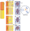
Basic model illustrating the cohesive role of dopa-Fe complexes in the byssus cuticle. Granules contain a higher cross-link density than matrix. When the cuticle is stretched to less than 30% strain, the randomly coiled mfp-1 chains begin to unravel, and the granule and matrix deform equivalently. However, when stretched beyond 30% strain mfp-1 chains are largely unraveled, and microcracks form outside the granules because of the difference in cross-link density. When relaxed, the granule returns to its initial shape, whereas microcracks do not exhibit immediate recovery.

Comment in

Similar articles

Cited by

References

    1. Broomell CC, Mattoni MA, Zok FW, Waite JH. J. Exp. Biol. 2006;209:3219. - PMC - PubMed
    1. Werneke SW, Swann C, Farquharson LA, Hamilton KS, Smith AM. J. Exp. Biol. 2007;210:2137. - PubMed
    1. Harrington MJ, Waite JH. Biomacromolecules. 2008;9:1480. - PubMed
    1. Harrington MJ, Gupta HS, Fratzl P, Waite JH. J. Struct. Biol. 2009;167:47. - PMC - PubMed
    1. Lee SM, et al. Science. 2009;324:488. - PubMed

Publication types