. 2010 May;298(5):G722-31.
doi: 10.1152/ajpgi.00387.2009. Epub 2010 Mar 4.

Effect of indomethacin on bile acid-phospholipid interactions: implication for small intestinal injury induced by nonsteroidal anti-inflammatory drugs

Affiliations

Effect of indomethacin on bile acid-phospholipid interactions: implication for small intestinal injury induced by nonsteroidal anti-inflammatory drugs

Yong Zhou et al. Am J Physiol Gastrointest Liver Physiol. 2010 May.

Abstract

The injurious effect of nonsteroidal anti-inflammatory drugs (NSAIDs) in the small intestine was not appreciated until the widespread use of capsule endoscopy. Animal studies found that NSAID-induced small intestinal injury depends on the ability of these drugs to be secreted into the bile. Because the individual toxicity of amphiphilic bile acids and NSAIDs directly correlates with their interactions with phospholipid membranes, we propose that the presence of both NSAIDs and bile acids alters their individual physicochemical properties and enhances the disruptive effect on cell membranes and overall cytotoxicity. We utilized in vitro gastric AGS and intestinal IEC-6 cells and found that combinations of bile acid, deoxycholic acid (DC), taurodeoxycholic acid, glycodeoxycholic acid, and the NSAID indomethacin (Indo) significantly increased cell plasma membrane permeability and became more cytotoxic than these agents alone. We confirmed this finding by measuring liposome permeability and intramembrane packing in synthetic model membranes exposed to DC, Indo, or combinations of both agents. By measuring physicochemical parameters, such as fluorescence resonance energy transfer and membrane surface charge, we found that Indo associated with phosphatidylcholine and promoted the molecular aggregation of DC and potential formation of larger and isolated bile acid complexes within either biomembranes or bile acid-lipid mixed micelles, which leads to membrane disruption. In this study, we demonstrated increased cytotoxicity of combinations of bile acid and NSAID and provided a molecular mechanism for the observed toxicity. This mechanism potentially contributes to the NSAID-induced injury in the small bowel.


Figures
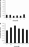
Fig. 1.
Fig. 1.
A: effect of indomethacin (Indo) alone on the permeability of gastric AGS cell plasma membrane. Cells were incubated in Ham's medium containing various concentrations of Indo for 3 h. Level of cytoplasmic enzyme lactate dehydrogenase (LDH) in the medium, an indication of cell plasma membrane leakage, was measured. The LDH level is here quantified by the absorbance level at wavelength of 490 nm normalized by subtracting absorbance at 690 nm, which were plotted against Indo concentrations. B: effect of Indo on cell viability measured by the level of 3-(4,5-dimethylthiazol-2-yl)-2,5-diphenyl tetrazolium bromide (MTT) incorporated into cells. After Indo treatment, medium containing Indo was discarded and cells were incubated in fresh medium containing 0.5% MTT for 2 h. MTT in the cells was then dissolved in MTT-solvent mix and MTT levels were quantified as absorbance at 570 nm, which were plotted against Indo concentrations.
Fig. 2.
Fig. 2.
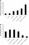
A: dose effect of Indo on the ability of deoxycholic acid (DC) to alter LDH level found in the medium, an indicator of membrane permeability. *Statistical significance vs. either 0.3 mM DC or 2 mM Indo alone. B: dose effect of Indo on the ability of DC to alter gastric AGS cell number. *Statistical significance vs. either 0.3 mM DC or 2 mM Indo alone. ctl, Control.
Fig. 3.
Fig. 3.
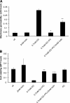
A: detrimental effect of Indo on the protective effect of purified soy phosphatidylcholine (PC) on cell plasma membrane permeability as indicated by LDH levels found in the medium. *Statistical significance vs. 0.7 mM DC; **statistical significance vs. 0.7 mM DC + 2.8 mM PC. B: ability of Indo to diminish the protective effect of PC on cell viability as indicated by levels of MTT incorporated into cells. *Statistical significance over control; **statistical significance vs. 0.7 mM DC; ***statistical significance vs. 0.7 mM DC + 2.8 mM PC.
Fig. 4.
Fig. 4.
Combined effect of Indo and conjugated bile acids (BA), taurodeoxycholic acid (TDC) and glycodeoxycholic acid (GDC), on intestinal epithelial IEC-6 cell plasma membrane permeability and cell viability. Similar effects were found as in AGS cells. IEC-6 cell plasma membrane permeability measured by LDH level in the cell medium was significantly increased by combining TDC and Indo (A) or GDC and Indo (B) compared with treatments with these agents alone. Cell viability measured by MTT incorporation into cells was markedly decreased by the combination of TDC and Indo (C) or GDC and Indo (D). A bile acid concentration of 0.3 mM was used in all bile acid-Indo combined treatments.
Fig. 5.
Fig. 5.
The amount of encapsulated calcein that permeated out of dilinoeoyl phosphatidylcholine (DLPC) liposomes was significantly increased in the presence of both DC and Indo, indicating the ability of a combination of DC and Indo to destabilize PL membranes. *Statistical significance vs. 0.4 mM DC + Indo.
Fig. 6.
Fig. 6.
DC and Indo dose dependently decreased the membrane dipole potential, indicating that the combination of DC and Indo negatively affects the packing density of DLPC membrane and thereby integrity.
Fig. 7.
Fig. 7.
Indomethacin (Indo) was added to a mixture of DC and dilinoleoyl PC (DLPC) liposomes. Energy transfer between bile acid and PC was weakened in the presence of Indo. FRET, fluorescence resonance energy transfer.
Fig. 8.
Fig. 8.
Zeta (ζ) potential of purified soy PC liposomes was measured in the presence of either DC alone, Indo alone, or the combination of both. Both DC and Indo alone caused the PC membrane to become negatively charged since both amphiphiles are weak acids and possess negative charge at neutral pH. The ζ-potential of PC bilayer exposed to both amphiphiles was found to be approximately the sum of the ζ-potential of PC membrane exposed to either amphiphile alone, indicating the association of both types of molecules with PC membrane. **Statistical significance vs. PC + 0.5 mM DC.
Fig. 9.
Fig. 9.
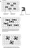
A: schematic demonstrating bile acid monomers partitioning into a phospholipid bilayer. A typical bile acid molecule possesses 2 faces: a hydrophilic face with hydroxyl groups attached and a hydrophobic face with the cholesterol backbone (see right). A top view of a lipid bilayer (lipids depicted by open circles) containing bile acids illustrates that 2 bile acid monomers form a dimer with the hydrophilic faces of the bile acid molecules hidden inside the dimer complex while exposing the hydrophobic face to the bilayer core. This reverse micelle structure ensures that the highly polar hydrophilic face of the bile acid would not interact unfavorably with the nonpolar bilayer core. A similar packing pattern is also found in PC-bile acid mixed micelles. B: amphiphilic Indo molecules (solid hexagons) potentially interact extensively with the hydrophilic face of the bile acid molecules and further expand the area that is polar. The original dimer structure becomes inadequate in protecting the polar portion of the bile acid/Indo molecules. C: unfavorable interactions between now-exposed hydrophilic face of the bile acid molecules and nonpolar bilayer core allows association of several bile acid and Indo molecules to form a larger complex. This change potentially further isolates bile acid molecules from PC lipids. Formation of the “polar pocket” composed of bile acids and Indo molecules induces increase in membrane permeability and disruption of the entire membrane. This is thought to occur when bile acid concentration is either below or above its critical micellar concentration.

Similar articles

Cited by

References

    1. Bjarnason I, Hayllar J, MacPherson AJ, Russell AS. Side effects of nonsteroidal anti-inflammatory drugs on the small and large intestine in humans. Gastroenterology 104: 1832–1847, 1993 - PubMed
    1. Brune K, Nurnberg B, Szelenyi I. The Enterohepatic Circulation of Some Anti-Inflammatory Drugs May Cause Intestinal Ulcerations Boston, MA: MTP Press, 1985
    1. Carey MC, Montet JC, Phillips MC, Armstrong MJ, Mazer NA. Thermodynamic and molecular basis for dissimilar cholesterol-solubilizing capacities by micellar solutions of bile salts: cases of sodium chenodeoxycholate and sodium ursodeoxycholate and their glycine and taurine conjugates. Biochemistry 20: 3637–3648, 1981 - PubMed
    1. Carey MC, Small DM. Micellar properties of dihydroxy and trihydroxy bile salts: effects of counterion and temperature. J Colloid Interface Sci 31: 382–396, 1969 - PubMed
    1. Clarke RJ. Effect of lipid structure on the dipole potential of phosphatidylcholine bilayers. Biochim Biophys Acta 1327: 269–278, 1997 - PubMed

Publication types

MeSH terms