

doi: 10.1093/nar/gkq133.
Epub 2010 Mar 4.
The crystal structure of unmodified tRNAPhe from Escherichia coli
Affiliations
- PMID: 20203084
- PMCID: PMC2896525
- DOI: 10.1093/nar/gkq133
Item in Clipboard
The crystal structure of unmodified tRNAPhe from Escherichia coli
Nucleic Acids Res.
2010 Jul.
Abstract
Post-transcriptional nucleoside modifications fine-tune the biophysical and biochemical properties of transfer RNA (tRNA) so that it is optimized for participation in cellular processes. Here we report the crystal structure of unmodified tRNA(Phe) from Escherichia coli at a resolution of 3 A. We show that in the absence of modifications the overall fold of the tRNA is essentially the same as that of mature tRNA. However, there are a number of significant structural differences, such as rearrangements in a triplet base pair and a widened angle between the acceptor and anticodon stems. Contrary to previous observations, the anticodon adopts the same conformation as seen in mature tRNA.
Figures
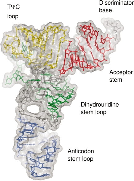
Structure of unmodified E. coli tRNAPhe. The nucleotides of the acceptor stem (red), D stem loop (green), ASL (blue) and TΨC loop (yellow) are coloured according to the region they belong to. Molecular surface is shown in grey.
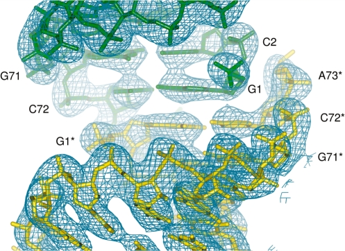
Crystal packing of the acceptor stem. G1–C72 base pairs from two tRNA molecules (yellow and green) are related by a crystallographic 2-fold axis. Nucleotides in one of the symmetry mates (yellow) are denoted with a star. Corresponding 2m|Fo|−D|Fc| electron density maps are contoured at 1.5σ.
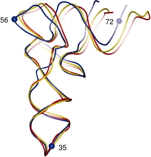
Difference in conformation between unmodified and modified tRNAs. Superposition of unmodified E. coli tRNAPhe (blue) and yeast tRNAPhe in its monoclinic form (yellow, PDB 1EHZ) and orthorhombic form (red, PDB 4TRA). The P atoms of residues 35, 56 and 72 (blue spheres) were used for the calculation of inter-arm angles.
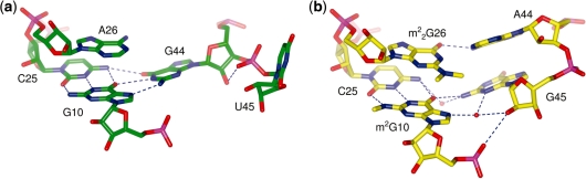
The rearrangement of triplet base pairs. (a) The triplet base pair in the unmodified E. coli tRNAPhe compared with (b) the triplet base pair in yeast tRNAPhe (PDB 1EHZ). Nucleotides are shown as sticks with oxygen atoms in red, nitrogen atoms in blue and phosphorus atoms in magenta. Water molecules are shown as red spheres and hydrogen bonds are depicted as blue dashed lines.
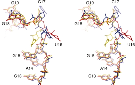
Conformation of the D-loop. Stereo view of the structures of unmodified E. coli tRNAPhe (blue) and yeast tRNAPhe in its monoclinic (yellow, PDB 1EHZ) and orthorhombic (red, PDB 4TRA) forms. In yeast tRNAPhe dihydrouridine is present at positions 16 and 17.
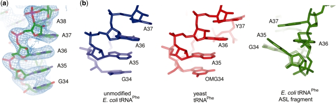
Anticodon stem loop conformation. (a) Electron density map for nucleotides 34–38. The 2m|Fo|−D|Fc| map is contoured at 1.5σ. (b) The conformation of these nucleotides (left) is essentially the same as in modified tRNA (middle) but differs from the conformation observed in the lowest energy conformer of the unmodified ASL fragment (right). All three ASLs are shown in the same orientation after superposition using the backbone P and C1′ atoms of nucleotides 31–39.
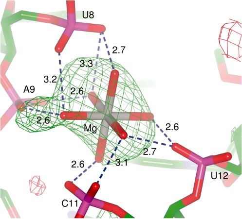
The coordination of the hydrated Mg2+ ion. The m|Fo|−D|Fc| electron density map contoured at 3σ was calculated after omitting the coordinating water molecules. The hydrogen bonds between the phosphate groups and the water molecules are depicted by dashed blue lines, along with the distances in Angstroms.
References
-
- Schurer H, Schiffer S, Marchfelder A, Morl M. This is the end: processing, editing and repair at the tRNA 3′-terminus. Biol. Chem. 2001;382:1147–1156. - PubMed
-
- Robertus JD, Ladner JE, Finch JT, Rhodes D, Brown RS, Clark BFC, Klug A. Structure of yeast phenylalanine transfer RNA at 3 Å resolution. Nature. 1974;250:546–551. - PubMed
Publication types
MeSH terms
Substances
Associated data
- Actions
