. 2010 Jul;24(7):2254-61.
doi: 10.1096/fj.09-137380. Epub 2010 Mar 4.

Crucial role of the small GTPase Rac1 in insulin-stimulated translocation of glucose transporter 4 to the mouse skeletal muscle sarcolemma

Affiliations

Crucial role of the small GTPase Rac1 in insulin-stimulated translocation of glucose transporter 4 to the mouse skeletal muscle sarcolemma

Shuji Ueda et al. FASEB J. 2010 Jul.

Abstract

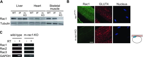
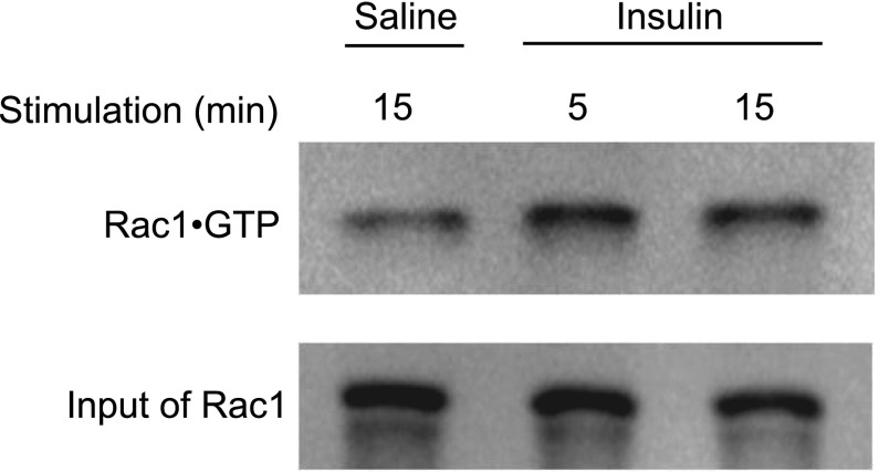
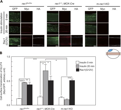
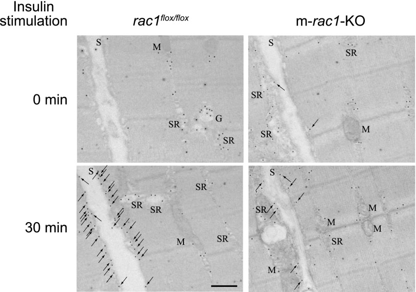
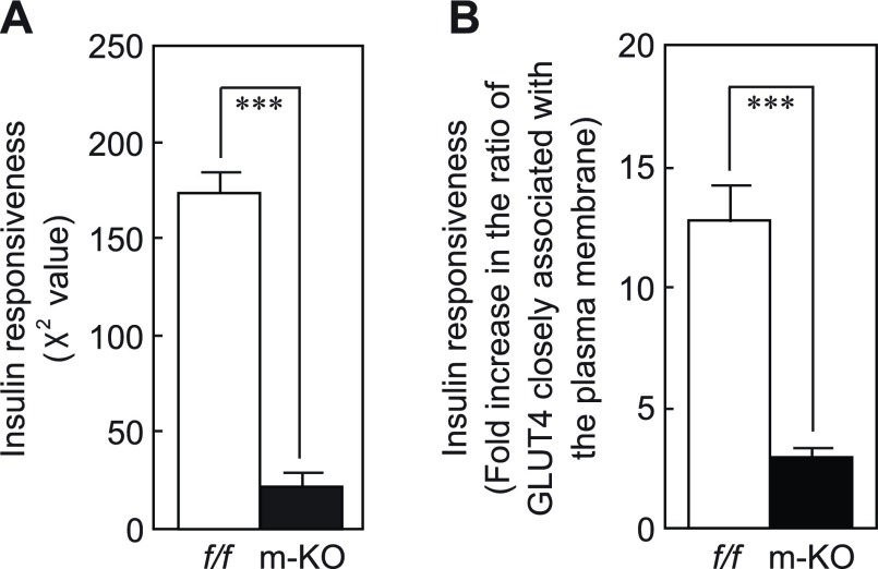
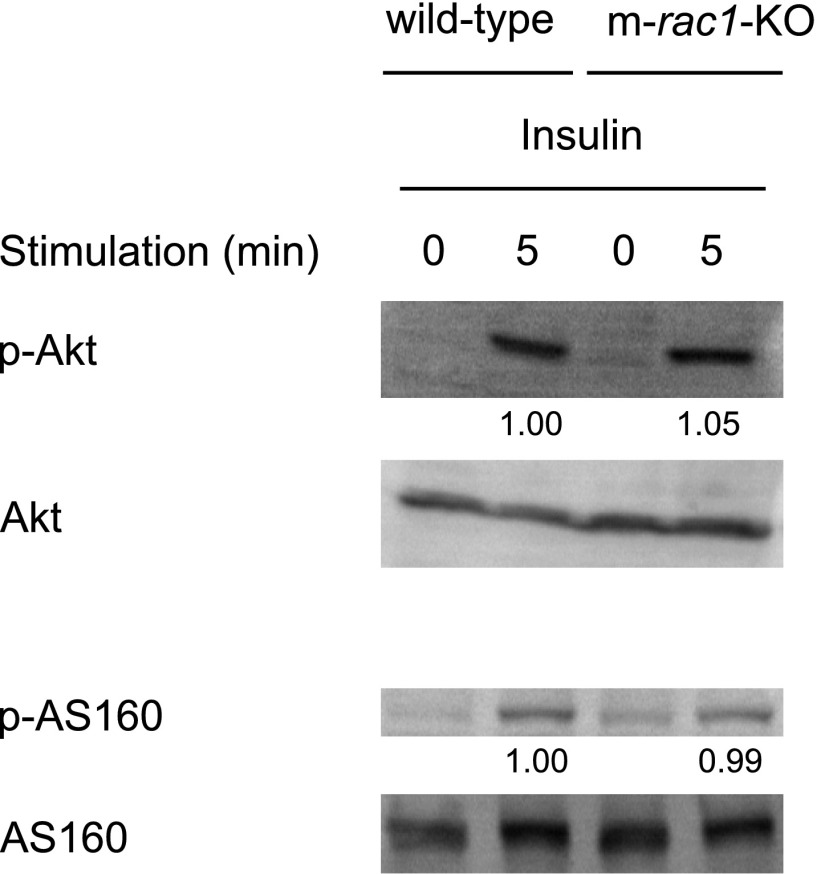
The Rho family GTPase Rac1 has been implicated in the regulation of glucose uptake in myoblast cell lines. However, no evidence for the role of Rac1 has been provided by a mouse model. The purpose of this study is to test the involvement of Rac1 in insulin action in mouse skeletal muscle. Intravenous administration of insulin indeed elicited Rac1 activation in gastrocnemius muscle, suggesting the involvement of Rac1 in this signaling pathway. We then examined whether insulin-stimulated translocation of the facilitative glucose transporter GLUT4 from its storage sites to the skeletal muscle sarcolemma depends on Rac1. We show that ectopic expression of constitutively activated Rac1, as well as intravenous administration of insulin, caused translocation of GLUT4 to the gastrocnemius muscle sarcolemma, as revealed by immunofluorescent staining of a transiently expressed exofacial epitope-tagged GLUT4 reporter. Of particular note, insulin-dependent, but not constitutively activated Rac1-induced, GLUT4 translocation was markedly suppressed in skeletal muscle-specific rac1-knockout mice compared to control mice. Immunogold electron microscopic analysis of endogenous GLUT4 gave similar results. Collectively, we propose a critical role of Rac1 in insulin-dependent GLUT4 translocation to the skeletal muscle sarcolemma, which has heretofore been predicted solely by cell culture studies.


Figures

Figure 1.
Figure 1.
Expression of Rac1 and GLUT4 in skeletal muscle. A) Expression level of Rac1 in liver, heart, and skeletal muscle (gastrocnemius muscle) of wild-type (WT), rac1flox/flox (f/f), and m-rac1-KO (m-KO) mice (8–10 wk old) was analyzed by immunoblotting with an anti-Rac1 antibody. Expression level of tubulin was also analyzed as a loading control. Ten micrograms of protein was loaded on each lane. B) Expression level and subcellular localization of Rac1 and GLUT4 in gastrocnemius muscle fibers of rac1flox/flox and m-rac1-KO mice (8–10 wk old) were analyzed by immunofluorescent staining with specific antibodies. Nuclei were stained with 4′,6-diamidino-2-phenylindole. Images were acquired from the focal plane, as depicted in schematic diagram at right. Blue ellipse in the schematic diagram indicates a nucleus. Scale bar = 20 μm. C) Expression of 3 Rac isoforms (Rac1, Rac2, and Rac3) in gastrocnemius muscle of wild-type and m-rac1-KO mice (8–10 wk old) was analyzed by RT-PCR.
Figure 2.
Figure 2.
Activation of Rac1 in insulin-stimulated mouse skeletal muscle. Insulin (5 IU/kg body wt) was administered intravenously to wild-type mice (8–10 wk old). Following stimulation with insulin for 5 or 15 min, Rac1·GTP level in soleus and gastrocnemius muscle was analyzed by pulldown assay.
Figure 3.
Figure 3.
Translocation of GLUT4 to the plasma membrane in skeletal muscle fibers, as revealed by immunofluorescent staining of an exofacial epitope-tagged GLUT4 reporter. A) GLUT4myc7-GFP was expressed with or without constitutively activated HA-tagged Rac1(G12V) in gastrocnemius muscle fibers of rac1flox/flox, rac1+/+; MCK-Cre, and m-rac1-KO mice (8–10 wk old). Insulin (5 IU/kg body wt) was administered intravenously. Total GLUT4myc7-GFP was visualized by fluorescence of GFP (green). GLUT4myc7-GFP translocated to the sarcolemma and externalized to the cell surface and Rac1(G12V) were visualized by immunofluorescent staining with anti-Myc (red) and anti-HA (white) antibodies, respectively. Images were acquired from the focal plane, as depicted in schematic diagram at bottom. Blue ellipse indicates a nucleus. Scale bar = 20 μm. B) Quantification of green and red fluorescence intensity in a representative field of a gastrocnemius muscle fiber, as shown in A. Data represent mean ± se ratios of fluorescence intensity for 3 to 9 independent experiments. *P < 0.05; **P < 0.01; ***P < 0.001.
Figure 4.
Figure 4.
Subcellular distribution of GLUT4 in skeletal muscle fibers, as revealed by immunogold electron microscopy. Insulin (5 IU/kg body wt) was administered intravenously to rac1flox/flox and m-rac1-KO mice (8–10 wk old), and subcellular distribution of GLUT4 in gastrocnemius muscle fibers was analyzed by immunogold electron microscopic analysis. Arrows indicate GLUT4 labeling in sarcolemma. G, Golgi apparatus; M, mitochondrion; S, sarcolemma; SR, sarcoplasmic reticulum. Scale bar = 500 nm.
Figure 5.
Figure 5.
Inhibition of insulin-stimulated redistribution of GLUT4 in skeletal muscle fibers by conditional rac1 knockout. The χ2 value (A) and fold increase in ratio of GLUT4 closely associated with the plasma membrane (B) in rac1flox/flox (f/f) and m-rac1-KO (m-KO) mice (8–10 wk old) (see Supplemental Table 1) are shown as means ± se for 5 independent experiments. ***P < 0.001.
Figure 6.
Figure 6.
Effect of conditional rac1 knockout on insulin-stimulated phosphorylation of Akt and AS160. Insulin (5 IU/kg body wt) was administered intravenously to wild-type and m-rac1-KO mice (8–10 wk old). Following stimulation with insulin for 5 min, phosphorylation level of Akt and AS160 in gastrocnemius muscle was analyzed by immunoblotting with phospho-specific antibodies. Relative intensities of bands are shown below the image. Sixty micrograms of protein was loaded on each lane.

References

    1. Klip A., Pâquet M. R. (1990) Glucose transport and glucose transporters in muscle and their metabolic regulation. Diabetes Care 13, 228–243 - PubMed
    1. Saltiel A. R., Kahn C. R. (2001) Insulin signalling and the regulation of glucose and lipid metabolism. Nature 414, 799–806 - PubMed
    1. Biddinger S. B., Kahn C. R. (2006) From mice to men: insights into the insulin resistance syndromes. Annu. Rev. Physiol. 68, 123–158 - PubMed
    1. Taniguchi C. M., Emanuelli B., Kahn C. R. (2006) Critical nodes in signalling pathways: insights into insulin action. Nat. Rev. Mol. Cell. Biol. 7, 85–96 - PubMed
    1. Huang S., Czech M. P. (2007) The GLUT4 glucose transporter. Cell Metab. 5, 237–252 - PubMed

Publication types