. 2010 Apr;24(4):722-34.
doi: 10.1210/me.2009-0326. Epub 2010 Mar 4.

Activation of Rac1 is closely related to androgen-independent cell proliferation of prostate cancer cells both in vitro and in vivo

Affiliations

Activation of Rac1 is closely related to androgen-independent cell proliferation of prostate cancer cells both in vitro and in vivo

Takashi Kobayashi et al. Mol Endocrinol. 2010 Apr.

Abstract

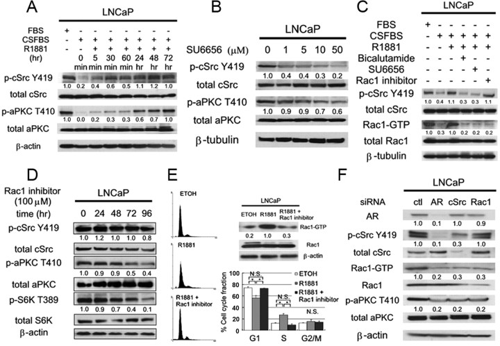
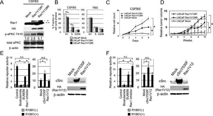
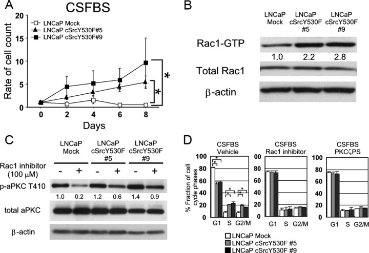
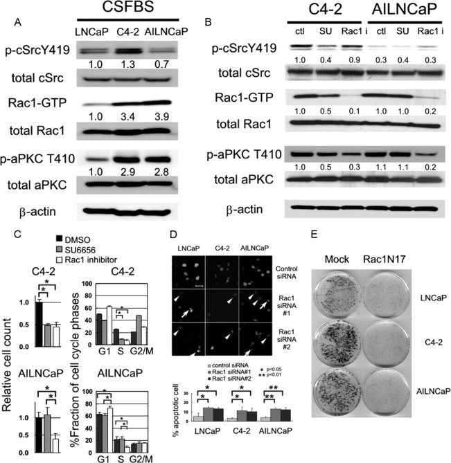
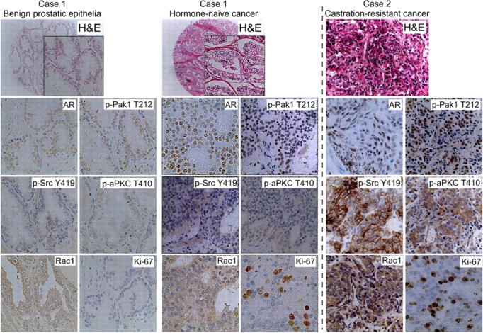
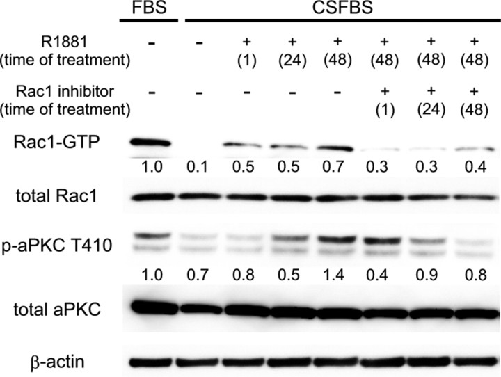
We and others previously showed that signaling through cSrc or atypical protein kinase C (aPKC) pathway regulates the proliferation of prostate cancer cells and is associated with their progression to castrate-resistance in vivo. However, the interrelation of these two kinases has been largely unexplored. In the present study, we show that androgen-induced activation of cSrc regulates the activity of aPKC through the small molecular weight G protein Rac1 in androgen-dependent LNCaP cells. Knockdown of cSrc in those cells reduces the phosphorylation of aPKC and the abundance of activated form of Rac1. Additionally, the treatment of those cells with Rac1 inhibitor repressed cell cycle progression at G(1)/S transition. In fact, forced expression of a constitutively active Rac1 mutant in LNCaP cells promoted cell proliferation under androgen-depleted conditions both in vitro and in vivo. Moreover, LNCaP C4-2 and AILNCaP cells, the syngeneic androgen-independent sublines from LNCaP cells, harbored abundant Rac1-GTP. Importantly, the inhibition of Rac1 suppressed cell proliferation and induced apoptotic cell death in all prostate cancer cell lines tested irrespective of their androgen-dependence. In immunohistochemical evaluation of tumor specimens from prostate cancer patients, Rac1 pathway appeared to be activated in the majority of castrate-resistant diseases. Collectively, our present results both in vitro and in vivo highly implicate that Rac1 can be a potential therapeutic target for patients with advanced prostate cancer, especially those with castrate-resistant status.


Figures

Fig. 1.
Fig. 1.
Androgen stimulation activates aPKC signals through cSrc via Rac1. The number below each band represents the mean value from densitometry reading. Densitometry readings normalized to total proteins are reported as averages from three independent experiments. A, LNCaP cells were left untreated or treated with CSFBS for 72 h followed by stimulation with 1 nm R1881 for indicated durations, and 60 μg of total cellular protein was separated by SDS-PAGE to examine indicated proteins by immunoblotting. B, LNCaP cells were treated with dimethylsulfoxide (DMSO) or SU6656 at indicated concentrations for 48 h, and total cell lysate containing 60 μg of protein was separated by SDS-PAGE to examine indicated proteins by immunoblotting. C, LNCaP cells were treated as indicated and cultured for 48 h. Then total cell protein was subjected to Rac1 pull-down assay or SDS-PAGE to examine indicated proteins by immunoblotting. D, LNCaP cells were treated with 100 μm Rac1 inhibitor for indicated durations (hr). Total cell lysate (60 μg) were separated by SDS-PAGE, and indicated proteins were examined by immunoblotting. E, LNCaP cells were cultured in phenol red-free RPMI 1640 supplemented with 10% CSFBS for 72 h and then treated ethanol or, R1881 (1 nm) with or without Rac1 inhibitor (100 μm) for 24 h. Cells were subjected to Rac1 pull-down assay and SDS-PAGE or flow cytometry analysis. Histograms represent triplicate independent experiments, and the column chart indicates the mean and sd values (*, P < 0.01). F, LNCaP cells were transfected with siRNA for indicated genes and harvested 48 h later for SDS-PAGE. The number below the band represents the mean value from densitometry reading.
Fig. 2.
Fig. 2.
LNCaP cells with forced expression of a constitutively active form of Rac1 (Rac1V12) acquire an ability of androgen-independent cell growth. LNCaP Mock and LNCaP Rac1V12 cells were incubated in RPMI 1640 supplemented with 10% FBS. Then, the medium was exchanged to RPMI 1640 supplemented with 10% CSFBS and were incubated for 72 h. A, Cells were harvested and 60 μg of total cellular protein was separated by SDS-PAGE. The indicated proteins were examined by immunoblotting. The number below each band represents the mean value from densitometry reading. Densitometry readings normalized to total proteins are reported as averages from two independent experiments. *, Exogenous HA-tagged Rac1; **, endogenous Rac1. B, Cell cycle was examined by flow cytometry analysis. Column chart indicates mean and sd (error bars) of them (*, P < 0.01). C, Cells were seeded into 6-cm dishes at 5.0 × 10⁵ cells/plate in RPMI 1640 supplemented with 10% FBS. After 24-h incubation for adhesion, cells on one dish were harvested, and the number of cells was counted as baseline (d 0). Then, the medium on other dish was exchanged to RPMI 1640 supplemented with 10% CSFBS, and the number of cells were counted on d 2, 4, 6, and 8. Results are shown as mean and sd of triplicate independent experiments (*, P < 0.01). D, Indicated cells were injected sc on a flank of male mice (n = 5 for each subline), and mice were surgically castrated after tumor formation was confirmed (see Materials and Methods). Then, the tumor volume was measured weekly. Results are shown as the mean and sd values (*, P < 0.01; bottom). E and F, Transactivity of endogenous AR in LNCaP (E) and exogeneous wt-AR in DU145 cells (F) was examined by dual-luciferase reporter assay (Promega). Cells were untreated (control) or treated with 10−6 m bicalutamide, 1 μm SU6656, or 100 μm Rac1 inhibitor, and the reporter activity was examined in the absence or presence of 10−11 m R1881 (left). Cells were transfected with pcDNA3.1(+) Mock, cSrcY530F, or HA-Rac1V12, and their effect on the reporter activity was examined in the absence or presence of R1881 (10−11 m) (right). In each column, relative luciferase activities to controls are shown with the sd values. Protein expressions under the same conditions for reporter assays were examined by Western blotting.
Fig. 3.
Fig. 3.
Activation of Rac1-aPKC pathway is required for androgen-independent proliferation of cultured LNCaP cSrcY530F cells. A, LNCaP Mock and LNCaP cSrcY530F cells were seeded into 6-cm dishes at 5.0 × 10⁵ cells/plate in RPMI 1640 supplemented with 10% FBS. After 24 h incubation for adhesion, cells on one dish were harvested, and the number of cells was counted as baseline (d 0). Then medium of the other dishes were replaced by phenol red-free RPMI 1640 supplemented with 10% CSFBS, and the number of cells was counted on the d 2, 4, 6, and 8. Results are shown as the mean and sd values of triplicate independent experiments (*, P < 0.001). B, Cells were treated with CSFBS for 72 h, and total protein was extracted. Rac1-GTP was separated by pull-down assay using 2 mg of total cellular protein and examined immunoblotting after SDS-PAGE. Other indicated proteins were separated by SDS-PAGE and examined by immunoblotting using 60 μg of total cellular protein. The number below each band represents the mean value from densitometry reading. Densitometry readings are normalized to total protein and reported as averages from two independent experiments. C, LNCaP Mock and cSrcY530F clones were cultured in CSFBS for 48 h and followed by the treatment with DDW or 100 μm Rac1 inhibitor for 48 h. Total cellular protein (60 μg) was separated by SDS-PAGE, and indicated proteins were examined by immunoblotting. The number below each band represents the mean value from densitometry reading. Densitometry readings are normalized to total protein and reported as averages from two independent experiments. D, Cell cycle was examined by flow cytometry analysis. Cells were seeded into six dishes at 5.0 × 10⁵ cells/plate in RPMI 1640 supplemented with 10% FBS. After 24 h incubation, medium was replaced by phenol red-free RPMI 1640 supplemented with 10% CSFBS. They were cultured for 24 h in the existence of 100 μm Rac1 inhibitor or 20 μm PKCζ pseudosubstrate inhibitor and harvested by trypsinization for flow cytometry. Column chart indicates the mean and sd values.
Fig. 4.
Fig. 4.
Activation of Rac1 is related to androgen-independent progression and survival of prostate cancer cells in culture. A, Total cellular protein (60 μg) of LNCaP, LNCaP C4-2, and AILNCaP cells were separated by SDS-PAGE, and indicated proteins were examined by immunoblotting. Rac1-GTP was separated by pull-down assay using 2 mg of total cellular protein and examined by immunoblotting after SDS-PAGE. The number below each band represents the mean value from densitometry reading. Densitometry readings are normalized to total protein and reported as averages from two independent experiments. B, C4-2 and AILNCaP cells were cultured in CSFBS and treated with dimethylsulfoxide (DMSO; ctl), SU6656 (1 μm) or Rac1 inhibitor (100 μm) 48 h before the extraction of total protein. Rac1-GTP was separated by pull-down assay with 2 mg of total cellular protein and examined immunoblotting after SDS-PAGE. Other indicated proteins were separated by SDS-PAGE and examined by immunoblotting with 60 μg of total cellular protein. The number below each band represents the mean value from densitometry reading. Densitometry readings are normalized to total protein and reported as averages from three independent experiments. C, C4-2 and AILNCaP cells were cultured in CSFBS and treated with DMSO, SU6656 (1 μm), or Rac1 inhibitor (100 μm) for 48 h before the harvest for flow cytometry. Column charts indicate the mean and sd values (*, P < 0.01). D, Cells were seeded into 6-cm dishes and transfected by 1 μg of pEGFP-N1 expressing H2B-enhanced GFP fusion protein alone or along with 12.5 ng of control (scramble) or Rac1 siRNA. Nuclear fragmentation (arrowheads) and chromatin condensation (arrows) were scored 48 h later using fluorescent microscopy. Column chart represents results of three independent experiments, in which at least 100 fluorescent cells were scored for chromatin condensation and nuclear fragmentation. Scale bar, 50 μm. E, LNCaP, C4-2 and AILNCaP cells were grown on 6-cm dishes, transfected with 8 μg pcDNA3.1(+) Mock (left dishes) or pcDNA3.1(+) HA-Rac1N17 (right dishes) with Lipofectamine 2000 reagent (Invitrogen) according to supplier’s protocol and cultured in RPMI 1640 medium supplemented with 10% FBS (LNCaP) or CSFBS (C4-2 and AILNCaP) containing 1 mg/ml of G418 for 2 wk. Colonies were visualized with 0.1% crystal violet staining.
Fig. 5.
Fig. 5.
Expression of AR, phospho-Src (Y419), phospho-Pak1 (T212), and phospho-aPKC (T410) in benign prostatic epithelial, hormone-naïve, and castrate-resistant human prostate cancer tissues. Representative immunohistochemical stainings of prostate cancer specimens from two individual patients are shown. Case 1, Gleason 3+3 hormone-naive prostate cancer, and adjacent benign prostatic epithelia obtained by radical prostatectomy undergone for prostate cancer as the primary treatment. Case 2, CRPC specimen obtained by transurethral resection for local relapse after androgen-deprivation therapy. Original magnification, ×200.
Fig. 6.
Fig. 6.
Effect of androgen stimulation and Rac1 inhibition on the phosphorylation levels of aPKC in LNCaP cells. LNCaP cells were grown in FBS or CSFBS flowed by stimulation with R1881 and/or Rac1 inhibitor. Total cellular protein was separated by SDS-PAGE, and then indicated proteins were examined by immunoblotting. Densitometry readings are normalized to total protein and reported as averages from two independent experiments.

Similar articles

Cited by

References

    1. Ferlay J, Bray F, Pisani P, Parkin D2001. Globocan 2000: cancer incidence, mortality and prevalence worldwide. Version 1.0. Lyon, France: IARC Press
    1. Jemal A, Siegel R, Ward E, Murray T, Xu J, Smigal C, Thun MJ2006. Cancer statistics, 2006. CA-Cancer J Clin 56:106–130 - PubMed
    1. Huggins C, Hodges CV1941. Studies on prostatic cancer: I. The effect of castration, of estrogen and of androgen injection on serum phosphatases in metastatic carcinoma of the prostate. Cancer Res 1:293–297 - PubMed
    1. Heinlein CA, Chang C2004. Androgen receptor in prostate cancer. Endocr Rev 25:276–308 - PubMed
    1. Unni E, Sun S, Nan B, McPhaul MJ, Cheskis B, Mancini MA, Marcelli M2004. Changes in androgen receptor nongenotropic signaling correlate with transition of LNCaP cells to androgen independence. Cancer Res 64:7156–7168 - PubMed

Publication types

MeSH terms