Review
. 2010 Nov;31(10):1778-86.
doi: 10.3174/ajnr.A2036. Epub 2010 Mar 4.

Lumbosacral transitional vertebrae: classification, imaging findings, and clinical relevance

Affiliations
Review

Lumbosacral transitional vertebrae: classification, imaging findings, and clinical relevance

G P Konin et al. AJNR Am J Neuroradiol. 2010 Nov.

Abstract

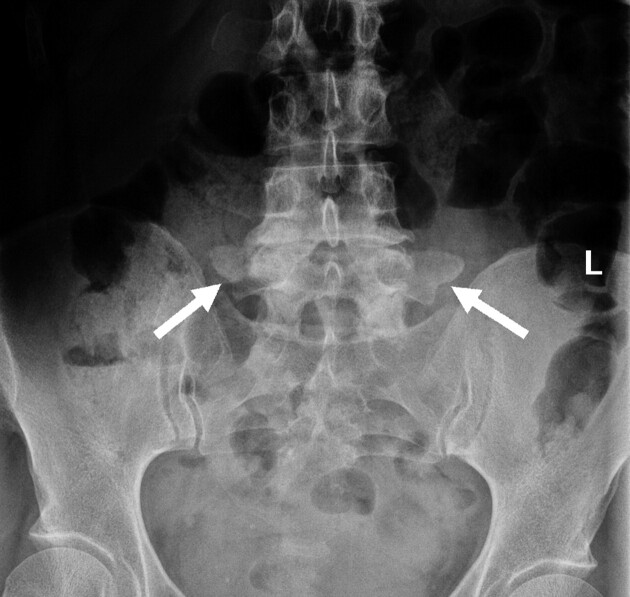
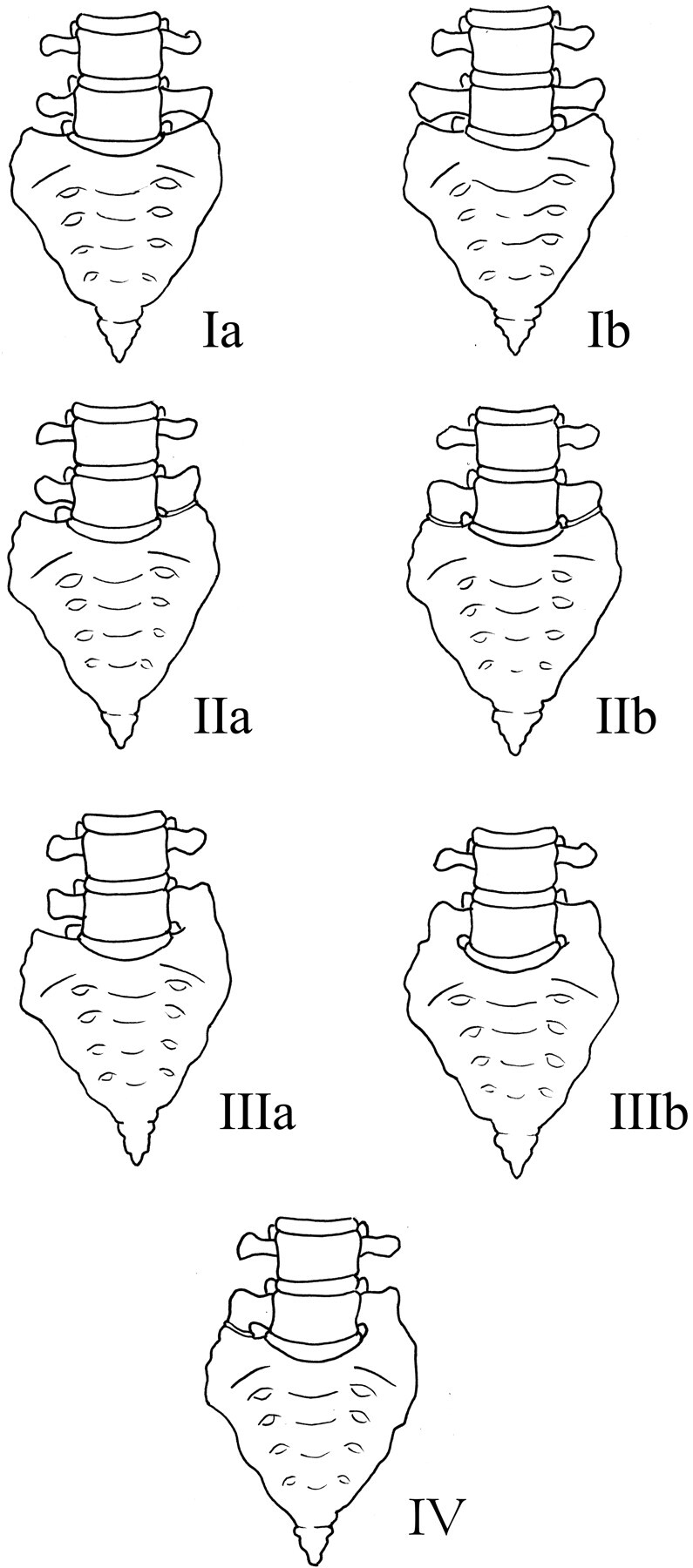
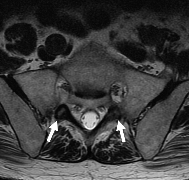
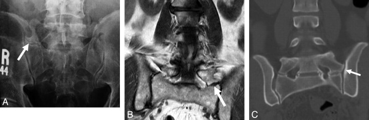
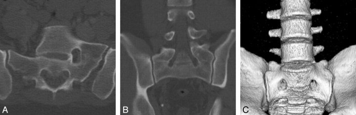
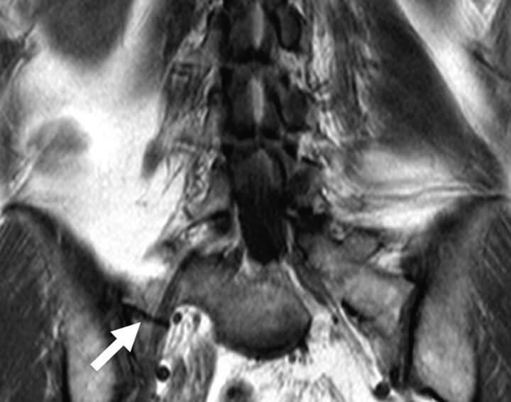
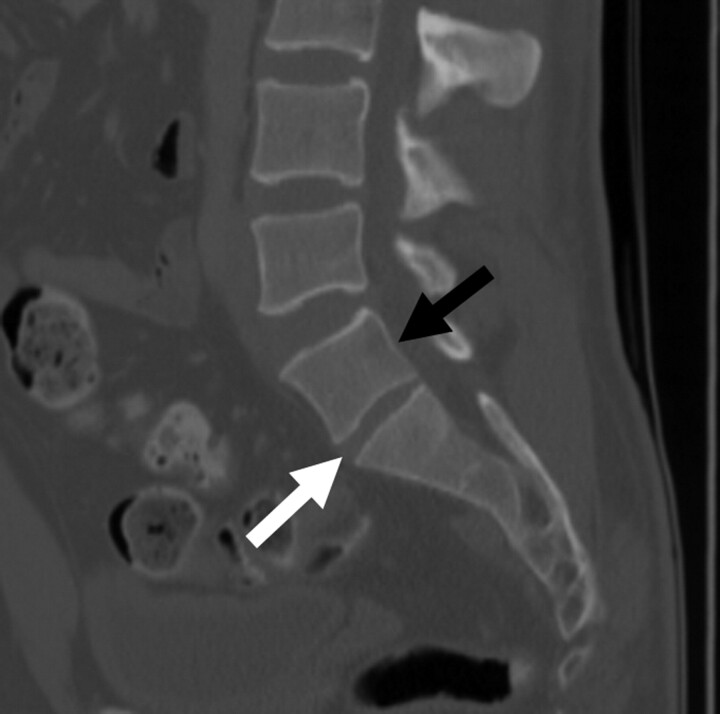
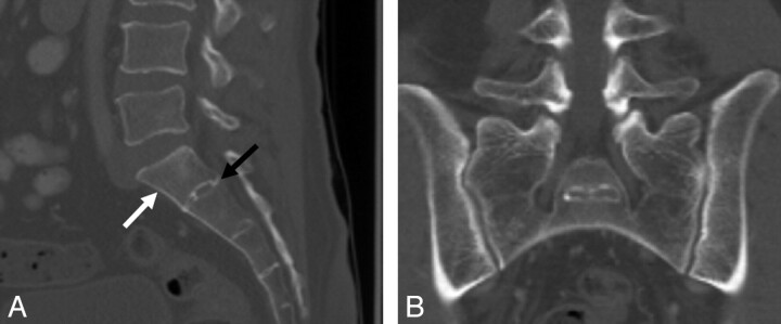
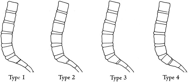
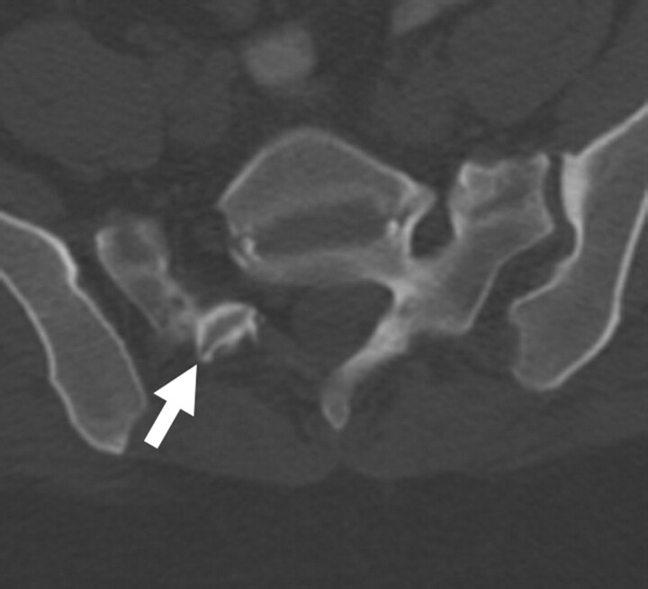
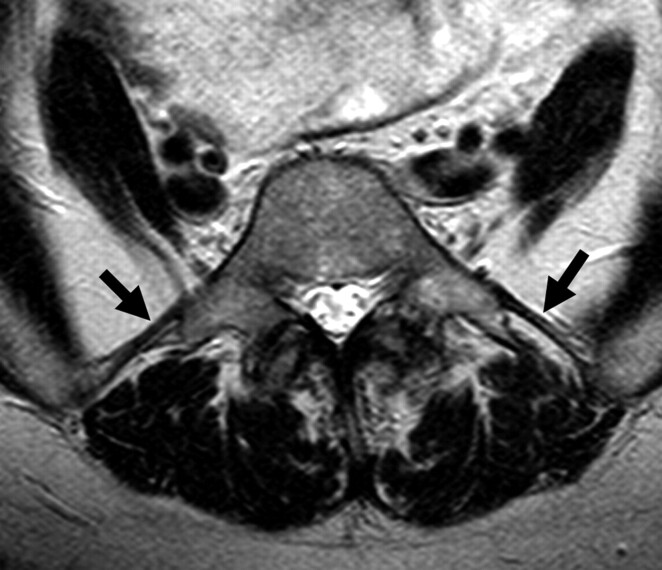
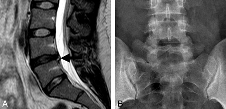
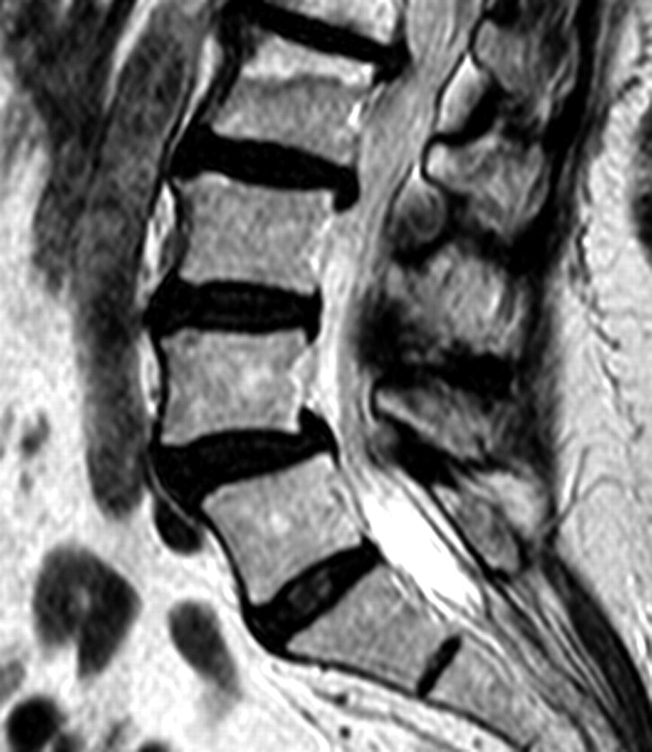
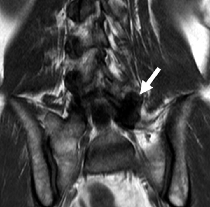
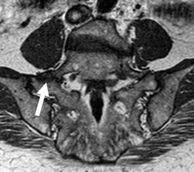
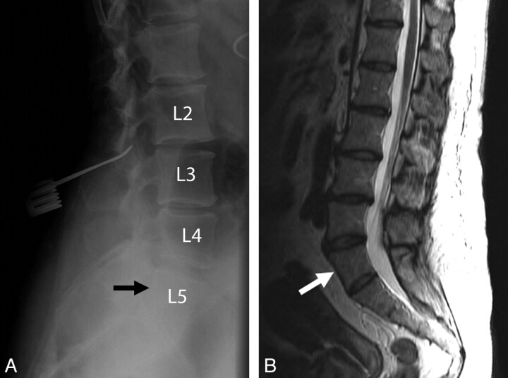
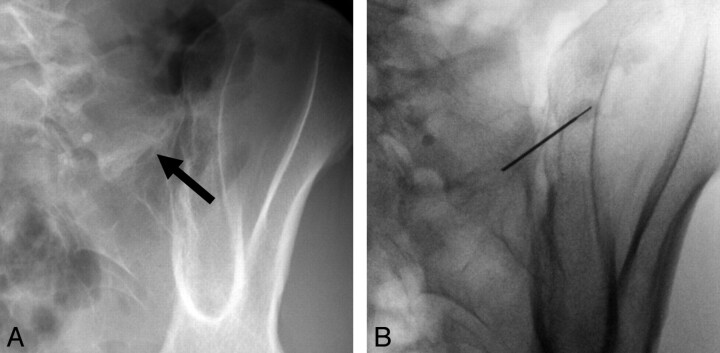
LSTVs are common within the spine, and their association with low back pain has been debated in the literature for nearly a century. LSTVs include sacralization of the lowest lumbar vertebral body and lumbarization of the uppermost sacral segment. These vertebral bodies demonstrate varying morphology, ranging from broadened transverse processes to complete fusion. Low back pain associated with an LSTV may arise from the level above the transition, the contralateral facet when unilateral, and/or the anomalous articulation when present. Although this association is still somewhat controversial, beyond dispute is the importance of identifying an LSTV in patients in whom a surgical or interventional procedure is planned. This is essential to avoid an intervention or surgery at an incorrect level. In this article, each of these issues will be addressed with attention to identifying and correctly numbering LSTVs as well as detecting imaging findings related to the genesis of low back pain.


Figures

Fig 1.
Fig 1.
Ferguson radiograph in a 35-year-old man. AP radiograph angled cranially at 30° allows better characterization of the transverse processes of L5. LSTVs have been classically described as best imaged by using Ferguson radiographs.
Fig 2.
Fig 2.
Illustration demonstrating the Castellvi classification of LSTVs.
Fig 3.
Fig 3.
Castellvi type Ib LSTV in a 53-year-old woman. AP radiograph demonstrates an LSTV with bilaterally enlarged L5 transverse processes (white arrows). There is no articulation with the sacrum.
Fig 4.
Fig 4.
Castellvi type IIa and IIb LSTVs. A, AP radiograph demonstrates an LSTV with a unilateral anomalous articulation of the right L5 transverse process with the sacrum (white arrow) in a 42-year-old man. B, T2-weighted coronal MR image demonstrates a unilateral anomalous articulation with the sacrum (white arrow) on the left in a 64-year-old man. C, Coronal reconstructed CT image demonstrates bilateral anomalous articulations of broadened transverse processes with the sacrum and the iliac bone on the left (white arrow) in a 52-year-old woman.
Fig 5.
Fig 5.
Castellvi type IIIa and IIIb LSTVs. A, Axial CT image demonstrates osseous fusion of the left L5 transverse process with the sacrum in a 36-year-old woman. B, Coronal reconstructed CT image demonstrates bilateral osseous fusion of L5 to the sacrum in a 31-year-old man. C, Volume-rendered CT image shows a Castellvi type IIIb LSTV with complete osseous fusion of the transverse processes to the sacrum in the same patient as seen in B.
Fig 6.
Fig 6.
Castellvi type IV LSTV in a 61-year-old woman. Coronal T2-weighted MR image demonstrates osseous fusion of the L5 transverse process to the sacrum on the left with an anomalous articulation on the right (white arrow).
Fig 7.
Fig 7.
Transitional S1 vertebral body in a 52-year-old woman. Sagittal CT image demonstrates “squaring” of a lumbarized S1 vertebral body (black arrow). Additionally, there is a fully-sized lumbar type disk between S1 and S2 (white arrow), compared with the characteristic vestigial disk typically seen at this level.
Fig 8.
Fig 8.
Transitional L5 vertebral body in a 52-year-old man. Sagittal CT image demonstrates “wedging” of a sacralized L5 transitional vertebral body (white arrow). A, Note the decreased height between the sacralized L5 vertebral body and S1 (black arrow) compared with the normal height typically seen at this level. B, Coronal CT image shows bilateral osseous fusion of the transverse processes of L5 with the sacrum (Castellvi type IIIb).
Fig 9.
Fig 9.
Illustration depicting the O'Driscoll classification system of S1–2 disk morphology.
Fig 10.
Fig 10.
Axial CT scan in a 52-year-old-woman with a Castellvi type IIa LSTV demonstrates fusion of the ipsilateral facet joint as well as a hypoplastic contralateral facet joint (white arrow) at L5-SI.
Fig 11.
Fig 11.
Axial T2-weighted MR image demonstrates rudimentary facet joints bilaterally (white arrows) at the S1-S2 level in this 79-year-old man with a lumbarized S1.
Fig 12.
Fig 12.
Iliolumbar ligaments in a 64-year-old woman. Axial T2-weighted MR image shows low-signal-intensity iliolumbar ligaments extending from the L5 transverse processes to the posteromedial iliac crests (arrows).
Fig 13.
Fig 13.
Adjacent-level disease. Sagittal T2-weighted MR image (A) demonstrates disk desiccation with a small central protrusion and associated annular tear (black arrow) in a 25-year-old female patient with a Castellvi type IIa LSTV as seen on this AP radiograph (B).
Fig 14.
Fig 14.
Adjacent-level disease in a 24-year-old woman. Sagittal T2-weighted MR image in a patient with a Castellvi type IIa LSTV demonstrates grade 1 anterolisthesis, disk desiccation, and disk bulging at the level above the transitional level.
Fig 15.
Fig 15.
Contralateral facet arthrosis in a 45-year-old man. Coronal T1-weighted MR image in a patient with a Castellvi type IIa LSTV demonstrates severe facet arthropathy (white arrow) contralateral to the anomalous articulation.
Fig 16.
Fig 16.
Degeneration of an anomalous articulation in an 84-year-old woman. Axial T1-weighted image demonstrates marked degeneration of the anomalous articulation on the right (white arrow) in a patient with a Castellvi type IIb LSTV.
Fig 17.
Fig 17.
Wrong-level spine surgery in a 55-year-old woman. A, Intraoperative radiograph demonstrates a localization device at what was believed to be the L3-L4 level based on misinterpretation of a sacralized L5 vertebral body (black arrow) as S1. B, Correlation with preoperative sagittal T2-weighted MR image better reveals the presence of the LSTV (white arrow) and, if used in combination with the intraoperative films, would have helped to avoid this surgical complication.
Fig 18.
Fig 18.
Injection of an anomalous articulation in a 48-year-old woman with right-sided low back pain and a Castellvi type IIa LSTV. A and B, Fluoroscopic spot images demonstrate degeneration of the anomalous articulation (arrow, A) and needle placement within the anomalous articulation for injection of anesthetic and corticosteroid (B).

References

    1. Bron JL, van Royen BJ, Wuisman PI. The clinical significance of lumbosacral transitional anomalies. Acta Orthop Belg 2007;73:687–95 - PubMed
    1. Castellvi AE, Goldstein LA, Chan DP. Lumbosacral transitional vertebrae and their relationship with lumbar extradural defects. Spine 1984;9:493–95 - PubMed
    1. Chang HS, Nakagawa H. Altered function of lumbar nerve roots in patients with transitional lumbosacral vertebrae. Spine 2004;29:1632–35 - PubMed
    1. Delport EG, Cucuzzella TR, Kim N, et al. . Lumbosacral transitional vertebrae: incidence in a consecutive patient series. Pain Physician 2006;9:53–56 - PubMed
    1. Elster AD. Bertolotti's syndrome revisited: transitional vertebrae of the lumbar spine. Spine 1989;14:1373–77 - PubMed

MeSH terms

LinkOut - more resources