. 2010 Apr;21(4):689-96.
doi: 10.1681/ASN.2009080829. Epub 2010 Mar 4.

Ultrastructural analysis of vascular calcifications in uremia

Affiliations

Ultrastructural analysis of vascular calcifications in uremia

Georg Schlieper et al. J Am Soc Nephrol. 2010 Apr.

Abstract

Accelerated intimal and medial calcification and sclerosis accompany the increased cardiovascular mortality of dialysis patients, but the pathomechanisms initiating microcalcifications of the media are largely unknown. In this study, we systematically investigated the ultrastructural properties of medial calcifications from patients with uremia. We collected iliac artery segments from 30 dialysis patients before kidney transplantation and studied them by radiography, microcomputed tomography, light microscopy, and transmission electron microscopy including electron energy loss spectrometry, energy dispersive spectroscopy, and electron diffraction. In addition, we performed synchrotron x-ray analyses and immunogold labeling to detect inhibitors of calcification. Von Kossa staining revealed calcification of 53% of the arteries. The diameter of these microcalcifications ranged from 20 to 500 nm, with a core-shell structure consisting of up to three layers (subshells). Many of the calcifications consisted of 2- to 10-nm nanocrystals and showed a hydroxyapatite and whitlockite crystalline structure and mineral phase. Immunogold labeling of calcification foci revealed the calcification inhibitors fetuin-A, osteopontin, and matrix gla protein. These observations suggest that uremic microcalcifications originate from nanocrystals, are chemically diverse, and intimately associate with proteinaceous inhibitors of calcification. Furthermore, considering the core-shell structure of the calcifications, apoptotic bodies or matrix vesicles may serve as a calcification nidus.


Figures

Figure 1.
Figure 1.
Microcalcifications can be noticed in approximately half of the uremic iliac artery segments as visualized by light microscopy. (A and B) Sample without detectable calcification. (C through E) The von Kossa stain shows microcalcifications in the media. Some cells seem to be completely surrounded by microcalcifications (arrows in E).
Figure 2.
Figure 2.
Mineral deposits of variable size can be found in a uremic artery (longitudinal view) by use of x-ray imaging. (A) Radiograph showing a thin plate of mineralization (*) in the blood vessel wall, with occasional nearby mineralization foci (arrows). (B) Three-dimensional reconstruction of mineral deposits after microcomputed tomography of a uremic artery. A large mineralized area is apparent in addition to small mineralization foci (arrows). The insert shows a single x-ray microcomputed tomographic “slice” from this region used in the reconstructions.
Figure 3.
Figure 3.
Microcalcifications in uremic arteries are located extracellularly in the vicinity of collagen and vesicular structures as shown by light and electron micrographs. (A) In undecalcified arterial samples embedded in plastic, sectioned, and examined by light microscopy for mineral deposits by von Kossa staining, numerous small mineralization foci of variable size (black arrows) near collagen fibrils (light blue staining, white arrows) are observed throughout the arterial media. The inset shows a higher magnification of the collagen matrix and mineral deposits. (B) Transmission electron micrograph showing a vascular smooth muscle cell (VSMC) surrounded by multiple small mineralization foci (black arrows) in the extracellular matrix. The white arrows indicate collagen fibrils. The inset shows a higher magnification of mineralization foci and collagen. (C) Mineralization foci (black solid arrows) in the vicinity of vesicular structures (black dashed arrows) and collagen fibrils (white arrows). (D through F) Transmission electron micrographs of mineralization foci in the extracellular matrix showing various morphologies. In many cases, there is evidence of a concentric lamellar pattern of the mineral deposits. The white arrow in F indicates cross-sectioned collagen fibrils.
Figure 4.
Figure 4.
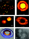
The diameter of microcalcifications ranged from 20 to 500 nm and a core-shell layered structure could be noticed in approximately one third of the microcalcifications which suggest that apoptotic bodies or matrix vesicles may serve as a calcification nidus. Microcalcifications seem to originate from 2- to 10-nm nanocrystals. (A) TEM showing multiple microcalcifications with diameters between 20 and 500 nm. Microcalcifications show various morphologies. A lamellar core-shell structure can be observed in many particles. (B through D) TEM of microcalcifications with a core-shell structure. A core was not present in all microcalcifications (D); however, when present (B and C), it consisted of more electron-dense material than the shell(s). (E) Three-dimensional reconstruction of the microcalcification shown in B with visualization of a solid core surrounded by an inner, less dense and an outer, more dense shell. (F) High-resolution TEM of a uremic vascular microcalcification. The microcalcifications consist of nanocrystals with a size of 2 to 10 nm.
Figure 5.
Figure 5.
Uremic arterial calcifications are chemically more diverse than previously thought with composition of hydroxyapatite and/or whitlockite. (A and B) Elemental and crystallographic analysis of microcalcifications using electron microscopy techniques. (A) EELS showing the spectrum of hydroxyapatite. (B) Electron diffraction pattern of hydroxyapatite. (C and D) Synchrotron radiation fluorescence and diffraction analysis. (C) Diffraction pattern for apatite (left) and whitlockite (right) standards. (D) The sample was scanned, and the results are depicted. The top panel shows the scan for the intensity of the calcium signal. The middle bottom panel shows the diffraction mapping, where the diffraction pattern of each point of the sample is represented in the scan on a one-to-one basis. The intensity of the signal is depicted by different colors. Both side panels show the diffraction pattern of the two points with the highest and second-highest calcium signal, respectively (as indicated by the white vertical line). The left spectrum reveals apatite as the chemical compound, whereas the right spectrum shows whitlockite.
Figure 6.
Figure 6.
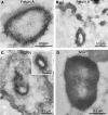
Calcification inhibitors are involved in the process of calcification in human arteries (high-resolution immunogold labeling of calcification foci revealed circulating and matrix mineral-binding proteins). Immunolabeling for fetuin-A (A and B), osteopontin (OPN; C), and matrix Gla protein (MGP; D) show strong to moderate gold particle labeling of mineralization foci in the uremic vessel wall. Immunolabeling in each case mainly localizes to electron-dense lamellar structures in the mineralization foci.

References

    1. Patient mortality and survival. United States Renal Data System. Am J Kidney Dis 32[ Suppl 1]: S69– S80, 1998 - PubMed
    1. Blacher J, Guerin AP, Pannier B, Marchais SJ, London GM: Arterial calcifications, arterial stiffness, and cardiovascular risk in end-stage renal disease. Hypertension 38: 938– 942, 2001 - PubMed
    1. Schlieper G, Kruger T, Djuric Z, Damjanovic T, Markovic N, Schurgers LJ, Brandenburg VM, Westenfeld R, Dimkovic S, Ketteler M, Grootendorst DC, Dekker FW, Floege J, Dimkovic N: Vascular access calcification predicts mortality in hemodialysis patients. Kidney Int 74: 1582– 1587, 2008 - PubMed
    1. Ketteler M, Westenfeld R, Schlieper G, Brandenburg V: Pathogenesis of vascular calcification in dialysis patients. Clin Exp Nephrol 9: 265– 270, 2005 - PubMed
    1. Moe SM, Reslerova M, Ketteler M, O'Neill K, Duan D, Koczman J, Westenfeld R, Jahnen-Dechent W, Chen NX: Role of calcification inhibitors in the pathogenesis of vascular calcification in chronic kidney disease (CKD). Kidney Int 67: 2295– 2304, 2005 - PubMed

Publication types