. 2010 Mar 3;30(9):3310-25.
doi: 10.1523/JNEUROSCI.5318-09.2010.

Functional organization of vestibular commissural connections in frog

Affiliations

Functional organization of vestibular commissural connections in frog

David Malinvaud et al. J Neurosci. .

Abstract

Central vestibular neurons receive substantial inputs from the contralateral labyrinth through inhibitory and excitatory brainstem commissural pathways. The functional organization of these pathways was studied by a multi-methodological approach in isolated frog whole brains. Retrogradely labeled vestibular commissural neurons were primarily located in the superior vestibular nucleus in rhombomeres 2/3 and the medial and descending vestibular nucleus in rhombomeres 5-7. Restricted projections to contralateral vestibular areas, without collaterals to other classical vestibular targets, indicate that vestibular commissural neurons form a feedforward push-pull circuitry. Electrical stimulation of the contralateral coplanar semicircular canal nerve evoked in canal-related second-order vestibular neurons (2 degrees VN) commissural IPSPs (approximately 70%) and EPSPs (approximately 30%) with mainly (approximately 70%) disynaptic onset latencies. The dynamics of commissural responses to electrical pulse trains suggests mediation predominantly by tonic vestibular neurons that activate in all tonic 2 degrees VN large-amplitude IPSPs with a reversal potential of -74 mV. In contrast, phasic 2 degrees VN exhibited either nonreversible, small-amplitude IPSPs (approximately 40%) of likely dendritic origin or large-amplitude commissural EPSPs (approximately 60%). IPSPs with disynaptic onset latencies were exclusively GABAergic (mainly GABA(A) receptor-mediated) but not glycinergic, compatible with the presence of GABA-immunopositive (approximately 20%) and the absence of glycine-immunopositive vestibular commissural neurons. In contrast, IPSPs with longer, oligosynaptic onset latencies were GABAergic and glycinergic, indicating that both pharmacological types of local inhibitory neurons were activated by excitatory commissural fibers. Conservation of major morpho-physiological and pharmacological features of the vestibular commissural pathway suggests that this phylogenetically old circuitry plays an essential role for the processing of bilateral angular head acceleration signals in vertebrates.


Figures

Figure 1.
Figure 1.
Photomicrographs of coronal sections showing confocal reconstructions of retrogradely labeled vestibular commissural, vestibulo-ocular, and vestibulo-spinal neurons. A–C, Application site of Alexa Fluor 488 dextran (green area) in the rostral region of the VN (A), crossing commissural axons in the midline (ml; inset in A) and retrogradely labeled cell bodies of commissural neurons in the contralateral VN (B, C); higher magnification (C) of labeled neurons in the outlined area in B. D, E, Separate populations of vestibular commissural neurons (green, red) after application of Alexa Fluor 488 dextran (green) into the rostral and of Alexa Fluor 546 dextran (red) into the caudal region of the vestibular nuclei. Only very few double-labeled cells (yellow) were labeled in the caudal region of the contralateral VN; higher magnification (E) of labeled neurons in the outlined area in D and in a section 100 μm more caudal (F). G–K, Target sites after application of Alexa Fluor 488 dextran (green) into the left VN (G) and of Alexa Fluor 546 dextran (red) into the right oculomotor nucleus (N.Oc; H). Separate populations of vestibular commissural (green) and vestibulo-ocular neurons (red) were retrogradely labeled in rostral regions of the VN (I–K); higher magnification (J) of labeled neurons in the outlined area in I and in a section 50 μm more caudal (K). L, M, Separate populations of retrogradely labeled vestibular commissural (green) and of vestibulo-spinal neurons (red) in caudal regions of the VN after application of Alexa Fluor 488 dextran into the VN and of Alexa Fluor 546 dextran bilaterally into the upper spinal cord; higher magnification (M) of labeled neurons in the outlined area in L. N, Commissural neurons in the DN after tracer application into the contralateral vestibular/auditory nuclei (see A) at the level of N.VIII; the inset shows DN neurons in N at higher magnification. White arrows in A, B, D, and L indicate the sulcus limitans. Scale bars: A, B, D, G, H, I, L, N, inset in A, 300 μm; C, E, J, M, inset in N, 100 μm; F, K, 50 μm. lat, Lateral; med, medial.
Figure 2.
Figure 2.
Schematic drawings of coronal sections through the dorsal hindbrain summarizing the location of vestibular/auditory commissural neurons. A–C, Location of retrogradely labeled neurons with respect to vestibular and auditory nuclei at five rostrocaudal levels after tracer applications into rostral (n = 6; A), intermediate (n = 6; B), and caudal (n = 5; C) areas of the contralateral vestibular nuclei. The different symbols in each row represent labeled cells from different experiments, respectively. The rostrocaudal level of the sections with respect to the caudal end of the entry of N.VIII in the brainstem (0 mm) is indicated in A; the rostrocaudal position of the sections with respect to the hindbrain segmental scaffold (r2–r7) was obtained from Straka et al. (2006) and is indicated in B. The outline of the vestibular subnuclei was adopted from Kuruvilla et al. (1985) and Matesz (1979). The arrow at each level in C indicates the sulcus limitans (s.l.).
Figure 3.
Figure 3.
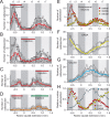
Distribution of vestibular commissural, GABAergic, and glycinergic vestibular neurons along the rostrocaudal extent of the vestibular nuclei. A–C, Number of labeled vestibular commissural neurons per section after application of Alexa Fluor 546 dextran (red bars) to rostral (A; n = 6), intermediate (B; n = 6), or caudal (C; n = 5) areas of the contralateral vestibular nuclei; different symbols in A–C represent individual experiments and the red line the average of these data, respectively. D, Number of double-labeled vestibular commissural neurons per section after application of Alexa Fluor 546 dextran to rostral (red bar) and Alexa Fluor 488 dextran (green bar) to caudal areas of the contralateral vestibular nuclei (n = 3); different symbols represent individual experiments and the orange line the average of these data. E, Average number of retrogradely labeled vestibular commissural neurons per section after rostral, intermediate, and caudal vestibular tracer applications (data from A–C). F–H, Average number of GABAergic (F) and glycinergic (G) vestibular neurons per section (data adopted from Reichenberger et al., 1997) and comparison with the normalized rostrocaudal distribution of retrogradely labeled vestibular commissural neurons (Com; H); different symbols in F and G represent individual experiments and the yellow (F) and blue line (G) the average of these data, respectively. In H, the number of cells per section was normalized to the maximal number of each type. Vertical stripes in the background of each plot indicate the hindbrain segmental scaffold (r2–r7) obtained from Straka et al. (2006). Note that GABAergic and vestibular commissural neurons have a similar rostrocaudal distribution between r2 and r6.
Figure 4.
Figure 4.
Photomicrographs of coronal sections showing confocal reconstructions of retrogradely labeled GABAergic or glycinergic commissural neurons. A–C, Combined GABA immunohistochemistry (red) and retrograde tracing with Alexa Fluor 488 dextran (green) revealed double-labeled GABA-immunopositive vestibular commissural neurons (yellow) in rostral regions of the VN (A, B) but not in the DN (C); higher magnification (B) of double-labeled vestibular commissural neurons (*) in the outlined area in A. Inset in B shows GABA-immunopositive but retrogradely unlabeled neurons in the VN from the adjacent section; retrogradely labeled commissural neurons in the DN (green) were GABA immunonegative but were surrounded by dense GABA-immunopositive terminal-like structures (red). Insets I and II show higher magnifications of DN neurons in the outlined area (II) and from the adjacent section (I). D–F, Combined glycine immunohistochemistry (red) and retrograde tracing with Alexa Fluor 488 dextran (green) revealed the absence of double-labeled glycine-immunopositive commissural neurons (yellow) in the VN (D, E) but moderate numbers in the DN (F). Higher magnification (E) of vestibular commissural (green) and of glycine-immunopositive neurons (red) in the outlined area in D; the inset in E shows glycine-immunopositive neurons in the VN from the adjacent section. Several retrogradely labeled (green) commissural neurons in the DN (F) were also glycine immunopositive (red) and thus appear yellow; the inset in F shows a higher magnification of the DN neurons in the outlined area. White arrows in A, C, D, and F indicate the sulcus limitans. Scale bars: A, C, D, F, 250 μm; B, E, insets in E, F, and inset II in C, 50 μm; inset in B and inset I in C, 20 μm.
Figure 5.
Figure 5.
Organization of brainstem commissural fiber pathways. A, Coronal section showing labeled vestibular and auditory fibers crossing the midline of the brainstem after application of Alexa Fluor 546 dextran to the right VN; the inset shows a coronal section with the application site in the VN at the level of N.VIII. B, Schematic drawing illustrating the organization of brainstem crossing commissural pathways between the bilateral vestibular and auditory nuclei (modified from Grofová and Corvaja, 1972); axons of commissural vestibular and auditory neurons cross the midline as an IAF and an EAF tract. C, Histological reconstructions of coronal sections at the level of N.VIII and iN.VIII-evoked afferent (black traces) and cN.VIII-evoked commissural responses (red traces) in 2°VN recorded after the lesion; depth and extension of longitudinal cuts of the bilateral EAF (C1), IAF (C2), and alternate EAF/IAF (C3) is indicated by the red areas. Stimulus intensities for the afferent responses ranged from 1.4 to 2.3 × T and for the commissural responses from 3.0 to 4.8 × T; records are the average of 20 responses, respectively, except for the orthodromic action potentials in C1 and C2, which are single sweeps. Arrowheads in C1–C3 indicate stimulus onset and dashed lines the resting membrane potential of −65 mV in C1, −66 mV in C2, and −64 mV in C3 on the left side and −67 mV in C3 on the right side. Calibration of responses to left iN.VIII and left cN.VIII in C3 apply also to responses to right iN.VIII and right cN.VIII. c, Contralateral; i, ipsilateral; s.l., sulcus limitans.
Figure 6.
Figure 6.
Synaptic organization of commissural inputs in phasic and tonic 2°VN. A, Schematic drawing illustrating the recording site in the right VN and the stimulation of individual semicircular canal nerve branches and the N.VIII on both sides. B, Commissural inputs in an identified phasic 2° AC neuron. B1, An initial subthreshold notch (black trace) and a single spike (gray trace) evoked by intracellular injection of a positive current step at two intensities (bottom traces) characteristic for phasic 2°VN; the inset in B1 shows the response onset at an extended timescale. B2, B3, iAC nerve-evoked monosynaptic afferent EPSP (B2) and cN.VIII-evoked disynaptic commissural EPSPs (B3); stimulus intensities in B2 (1.3, 2.0, 2.8 × T) and in B3 (2.1, 3.0, 4.0 × T). B4–B6, Separate stimulation of the three contralateral semicircular canal nerves evoked an oligosynaptic commissural IPSP from the coplanar cPC (B4) and disynaptic and oligosynaptic EPSPs from the two non-coplanar cAC (B5) and cHC (B6), respectively. Stimulus intensities are 2.5, 3.0, and 4.5 × T in B4, 2.2, 3.3, and 4.8 × T in B5, and 2.5, 3.8, and 5.0 × T in B6; note that all responses in B are from the same neuron. C, Coplanar semicircular canal commissural IPSPs in a tonic 2° PC neuron. C1, A continuous discharge in response to a positive current step (bottom trace) characteristic for tonic 2°VN. C2, Monosynaptic afferent EPSPs after electrical stimulation of the iPC nerve at 1.3 and 2.2 × T. C3, Disynaptic commissural IPSPs after stimulation of the coplanar cAC nerve at 2.2, 3.0, and 4.6 × T. D, Coplanar semicircular canal commissural EPSPs in a phasic 2° HC neuron. D1, A single spike in response to a positive current step (bottom trace) characteristic for phasic 2°VN. D2, Monosynaptic afferent EPSPs after electrical stimulation of the iHC nerve at 1.3 and 2.1 × T. D3, Commissural EPSPs after stimulation of the coplanar cHC nerve at 2.2, 3.0, and 4.6 × T with a disynaptic onset. E, Latency distribution of coplanar semicircular canal nerve-evoked commissural IPSPs (n = 138) and EPSPs (n = 51) in all recorded 2°VN; response latencies were distinguished into disynaptic (black bars) and oligosynaptic (>disynaptic, gray bars) onsets. Records in B1, C1, and D1 are single sweeps and in B2–B6, C2, C3, D2, and D3 the average of 20 responses. Arrowheads in B2–B6, C2, C3, D2, and D3 mark stimulus onset and dashed lines the resting membrane potential of the neurons, respectively; vertical gray bars in B3–B6, C3, and D3 indicate the mean ± SE of the disynaptic onset of the vestibular commissural field potential. Calibration in B2, and C2 apply to B3–B6 and C3, respectively. AC, Anterior canal; PC, posterior canal; HC, horizontal canal; c, contralateral; I, ipsilateral; CB, cerebellum; OT, optic tectum.
Figure 7.
Figure 7.
Parameters of coplanar semicircular canal commissural inputs in phasic and tonic 2°VN. A, All-or-nothing axonal action potentials recorded in the vestibular nuclei after electrical stimulation of the cAC nerve at threshold intensity; the inset shows the onset at an extended timescale. The onset latency is compatible with presynaptic action potentials that evoke IPSPs in 2°VN with a disynaptic onset (dotted line, arrow). B, Coplanar semicircular canal commissural IPSPs in a tonic 2° PC neuron. B1, cAC nerve-evoked disynaptic commissural IPSPs at stimulus intensities of 2.1, 2.5, 3.2, 3.9, and 4.5 × T; stronger stimuli recruited longer-latency components (arrows). B2, Commissural cAC nerve-evoked (3.2 × T) IPSPs at different membrane potentials; changes in IPSP amplitude with membrane polarization were used to determine the reversal potential. B3, Monosynaptic EPSPs after iPC nerve stimulation at 1.2, 1.7, and 2.3 × T. B4, Continuous discharge in response to a positive current step (bottom trace) characteristic for tonic 2°VN; note that all responses in B are from the same neuron. C, Coplanar semicircular canal commissural IPSPs in a phasic 2° PC neuron. C1, Monosynaptic EPSP after iPC nerve stimulation at 1.6 × T. C2, Single spike discharge in response to a positive current step (bottom trace) characteristic for phasic 2°VN. C3, Commissural cAC nerve-evoked (4.5 × T) IPSPs at different membrane potentials; note that IPSPs did not reverse with membrane hyperpolarization. D, Relative proportion of commissural IPSPs and EPSPs in tonic and phasic 2°VN. E, Relative proportion of commissural IPSPs with disynaptic and oligosynaptic (>di) onsets in tonic and phasic 2°VN. F, Amplitude distribution of commissural IPSPs evoked with stimulus intensities of >4.5 × T in phasic and tonic 2°VN at resting membrane potential. G, Dependency of commissural IPSP amplitudes on membrane polarization in tonic and phasic 2°VN; IPSPs in tonic 2°VN reverse at approximately −74 mV (black dotted line, vertical arrow), whereas IPSPs in phasic 2°VN remain small and independent of the membrane potential (gray dashed line). Records in A, B4, and C2 are single sweeps and in B1–B3, C1, and C3 the average of 20 responses. Arrowheads in A, B1–B3, C1, and C3 mark stimulus onset and dashed lines the membrane potential, respectively; gray bars in B1, B2, and C3 indicate the mean ± SE of the disynaptic onset of the vestibular commissural field potential. Calibration in B1 applies to B2.
Figure 8.
Figure 8.
Characterization of the intrinsic response dynamics of vestibular commissural neurons. A, Schematic drawing illustrating possible commissural connections with respect to the intrinsic response dynamics of tonic (gray) and phasic (black) 2°VN; disynaptic commissural responses could be mediated by tonic (1), phasic (3), or both types of 2°VN (2). B, Typical responses of a tonic (B1) and a phasic (B2) 2°VN to stimulation of the iAC (B1) and iHC (B2) nerve with a train of single electrical pulses (at 2.9 × T in B1 and at 3.2 × T in B2) that were sinusoidally modulated in frequency between 0 and 70 Hz (bottom traces); normalized rate of evoked spike discharge (B3) during pulse trains with peak frequencies of 70 Hz in tonic (dashed line) and phasic (solid line) 2°VN (adopted from Pfanzelt et al., 2008). C, Commissural compound IPSP of a tonic 2° AC neuron after electrical stimulation of the coplanar cPC nerve by a sinusoidally modulated pulse train (at 4.0 × T) with a peak frequency of 70 Hz (bottom traces); the inset shows the IPSP to the first single pulse (gray area) of the train (open arrowhead) at an extended timescale. Double arrows indicate the activation of individual IPSPs by the last two single pulses of the train. D, Model compound commissural inhibition in a tonic 2°VN after sinusoidal pulse train stimulation at a peak frequency of 70 Hz (bottom traces) with the assumption that the response was mediated only by phasic (D1), only by tonic (D3), or by equal proportions of both types of 2°VN (D2); the parameters to generate the model responses were obtained from the commissural IPSPs of the neuron shown in C at a resting membrane potential of −67 mV (see also Materials and Methods); the arrowhead in D1 indicates the truncation of the model response by the termination of the presynaptic discharge of putative phasic commissural 2°VN. E, Commissural compound EPSP of a phasic 2° PC neuron after electrical stimulation of the coplanar cAC nerve by a sinusoidally modulated pulse train (at 3.8 × T) with a peak frequency of 40 Hz (bottom traces); double arrows indicate the activation of individual EPSPs by the last two single pulses. Records in B1 and B2 are single sweeps and in C the average of 10 responses; dashed lines indicate the resting membrane potential, respectively; Calibration in B1 and D3 apply to B2 and D1, D2, respectively. aff, Afferent; com, commissural; s.c., semicircular canal.
Figure 9.
Figure 9.
Pharmacological profiles of semicircular canal commissural responses in phasic and tonic 2°VN. A, cAC nerve-evoked commissural field potentials in the vestibular nuclei before (black traces; control), during subsequent application of 2 μm strychnine (red trace in A1) and 5 μm bicuculline (red trace in A2), and after washout (gray traces; wash). B–E, Effect of glycinergic and GABAergic antagonists on coplanar semicircular canal nerve-evoked commissural responses in four different 2°VN; neurons were identified as 2° semicircular canal neurons by monosynaptic EPSPs from the iHC (B1), iPC (C1), or iAC (D1, E1) nerves and characterized as tonic 2°VN by a continuous discharge (B2, C2, E2) or as phasic 2°VN by a single spike (D2) in response to intracellular injected positive current pulses (indicated by bottom gray traces: 0.5 nA in B3, E3; 0.4 nA in C3; 0.7 nA in D3). B3, C3, D3, Effect of 2 μm strychnine on semicircular canal nerve-evoked commissural inhibition (gray traces, respectively); strychnine leaves disynaptic responses unaffected but reduces longer-latency components (black arrows in B3, C3, D3). C3, D3, Effect of 5 μm bicuculline and 2 μm strychnine on semicircular canal nerve-evoked commissural responses (red traces, respectively); bicuculline blocks all IPSP components including those with a disynaptic onset (red arrow in C3) and unmasks or increases EPSPs with a disynaptic onset in phasic 2°VN (red arrow in D3). E3, Effect of 100 μm CGP 35348 (gray trace) and combined application of 100 μm CGP 35348 and 5 μm bicuculline (red trace) on semicircular canal nerve-evoked commissural responses; CGP 35348 slightly reduces the IPSP amplitude, whereas bicuculline in the presence of CGP 35348 blocks the disynaptic IPSP completely and unmasks an EPSP with a disynaptic onset (red arrow). F, Proportion of coplanar canal commissural IPSPs with a disynaptic (F1) or oligosynaptic onset (>di; F2) that were reduced by bicuculline (Bic), CGP 35348 (CGP), or strychnine (Stry), respectively. Numbers in F indicate recorded neurons. Records in B2, C2, D2, and E2 are single sweeps and in A1, A2, B1, B3, C1, C3, D1, D3, E1, E3 the average of 20 responses. Arrowheads in A, B1, B3, C1, C3, D1, D3, E1, E3 mark stimulus onset and dashed lines in A the baseline and in B–E the resting membrane potential. AC, Anterior canal; PC, posterior canal; HC, horizontal canal; c, contralateral; i, ipsilateral.
Figure 10.
Figure 10.
Summary diagram depicting the major organizational principles of the vestibular commissure. A, Top view of a schematic hindbrain (left side) illustrating the rostrocaudal arrangement of the vestibular nuclei along the rhombomeric scaffold (A1) and of distinct vestibular subgroups with unique projections to major vestibulo-motor-related targets (A2), suggesting a feedforward push–pull organization of the vestibular commissure. B, Physio-pharmacological organization of pathways mediating a coplanar semicircular canal commissural inhibition in tonic (T) 2°VN (B1) and a coplanar semicircular canal commissural excitation in phasic (P) 2°VN (B2). VC, Vestibulo-cerebellar neurons; VCom, vestibular commissural neurons; VN, vestibular nucleus; VO, vestibulo-ocular neurons; VS, vestibulo-spinal neurons. Green neurons are excitatory, red neurons are inhibitory, and orange neurons are excitatory or inhibitory.

Similar articles

Cited by

References

    1. Ariel M, Fan TX, Jones MS. Bilateral processing of vestibular responses revealed by injecting lidocaine into the eighth cranial nerve in vitro. Brain Res. 2004;999:106–117. - PubMed
    1. Babalian AL, Vidal PP. Floccular modulation of vestibuloocular pathways and cerebellum-related plasticity: an in vitro whole brain study. J Neurophysiol. 2000;84:2514–2528. - PubMed
    1. Babalian A, Vibert N, Assie G, Serafin M, Mühlethaler M, Vidal PP. Central vestibular networks in the guinea-pig: functional characterization in the isolated whole brain in vitro. Neuroscience. 1997;81:405–426. - PubMed
    1. Bagnall MW, Stevens RJ, du Lac S. Transgenic mouse lines subdivide medial vestibular nucleus neurons into discrete, neurochemically distinct populations. J Neurosci. 2007;27:2318–2330. - PMC - PubMed
    1. Beraneck M, Pfanzelt S, Vassias I, Rohregger M, Vibert N, Vidal PP, Moore LE, Straka H. Differential intrinsic response dynamics determine synaptic signal processing in frog vestibular neurons. J Neurosci. 2007;27:4283–4296. - PMC - PubMed

Publication types

MeSH terms

LinkOut - more resources