. 2010 Mar 3;30(9):3398-408.
doi: 10.1523/JNEUROSCI.5620-09.2010.

Striatal muscarinic receptors promote activity dependence of dopamine transmission via distinct receptor subtypes on cholinergic interneurons in ventral versus dorsal striatum

Affiliations

Striatal muscarinic receptors promote activity dependence of dopamine transmission via distinct receptor subtypes on cholinergic interneurons in ventral versus dorsal striatum

Sarah Threlfell et al. J Neurosci. .

Abstract

Striatal dopamine (DA) and acetylcholine (ACh) regulate motivated behaviors and striatal plasticity. Interactions between these neurotransmitters may be important, through synchronous changes in parent neuron activities and reciprocal presynaptic regulation of release. How DA signaling is regulated by striatal muscarinic receptors (mAChRs) is unresolved; contradictory reports indicate suppression or facilitation, implicating several mAChR subtypes on various neurons. We investigated whether mAChR regulation of DA signaling varies with presynaptic activity and identified the mAChRs responsible in sensorimotor- versus limbic-associated striatum. We detected DA in real time at carbon fiber microelectrodes in mouse striatal slices. Broad-spectrum mAChR agonists [oxotremorine-M, APET (arecaidine propargyl ester tosylate)] decreased DA release evoked by low-frequency stimuli (1-10 Hz, four pulses) but increased the sensitivity of DA release to presynaptic activity, even enhancing release by high frequencies (e.g., >25 Hz for four pulses). These bidirectional effects depended on ACh input to striatal nicotinic receptors (nAChRs) on DA axons but not GABA or glutamate input. In caudate-putamen (CPu), knock-out of M(2)- or M(4)-mAChRs (not M(5)) prevented mAChR control of DA, indicating that M(2)- and M(4)-mAChRs are required. In nucleus accumbens (NAc) core or shell, mAChR function was prevented in M(4)-knock-outs, but not M(2)- or M(5)-knock-outs. These data indicate that striatal mAChRs, by inhibiting ACh release from cholinergic interneurons and thus modifying nAChR activity, offer variable control of DA release probability that promotes how DA release reflects activation of dopaminergic axons. Furthermore, different coupling of striatal M(2)/M(4)-mAChRs to the control of DA release in CPu versus NAc suggests targets to influence DA/ACh function differentially between striatal domains.


Figures

Figure 1.
Figure 1.
Striatal mAChRs enhance activity dependence of dopamine release. a–d, Mean peak [DA]o ± SEM versus frequency during four pulse trains (1–100 Hz) in controls (filled circles) or Oxo-M (triangles) normalized to [DA]o released by a single pulse in control conditions (a, c) or each individual condition (b, d), in CPu (n = 9–12) (a, b) and NAc (n = 9–14) (c, d). Significant effects of frequency: Kruskal–Wallis, p < 0.001. Curve fits are Gaussian (controls, R2 > 0.7) or sigmoidal curves (Oxo-M, R2 > 0.99). Significance of post hoc t tests for comparisons of Oxo-M versus controls are indicated by asterisks (**p < 0.01; ***p < 0.001). e, g, Profiles of mean [DA]o ± SEM versus time in CPu after stimuli (arrows) of either a single pulse (1p) or a high-frequency burst (4p/100 Hz) in controls versus drug conditions (Oxo-M, 10 μm; atropine, 2 μm). Data are normalized to peak [DA]o released by 1p in controls. n = 9. f, h, Mean peak [DA]o ± SEM versus number of pulses at 100 Hz normalized to release by a single pulse in own condition in CPu. e, f, The enhanced sensitivity of DA release to burst versus nonburst stimuli in Oxo-M (***p < 0.001 vs control; post hoc Tukey's t tests; n = 9) was reversed by addition of atropine (†††p < 0.001, Oxo-M vs Oxo-M+atropine; post hoc Tukey's t tests; n = 9) to a ratio not different from controls. g, h, Atropine alone does not modify DA release (post hoc Tukey's t tests; n = 9) but prevents Oxo-M-induced change in DA release probability (post hoc Tukey's t tests; n = 9).
Figure 2.
Figure 2.
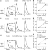
Muscarinic control of striatal DA release is via regulation of ACh tone at nicotinic AChRs. a, c, e, Profiles of mean [DA]o ± SEM versus time in CPu after stimuli (arrows) of either a single pulse (1p) or a high-frequency burst (4p/100 Hz). Data are normalized to peak [DA]o released by 1p in controls. Post hoc Bonferroni's t tests for burst versus single pulses are significant at p < 0.001 (n = 9). b, d, f, Mean peak [DA]o ± SEM versus number of pulses at 100 Hz in CPu normalized to release by a single pulse in own condition. a, b, DHβE causes profound changes in DA release but prevents additional effects of Oxo-M (b) (post hoc Tukey's t tests, ***p < 0.001 vs control, drug treatments not different from each other). c, d, DHβE is ineffective after previous oxotremorine-M application (d) (p > 0.05, post hoc Tukey's t tests, DHβE versus Oxo-M; ***p < 0.001 for comparisons with control). e, f, In contrast, a mixture of antagonists (Antags) for GABA (100 μm picrotoxin, 50 μm saclofen) and glutamate receptors [10 μm GYKI 52466, 50 μm d-AP5, 200 μm (S)-MCPG] does not prevent profound changes in DA release on subsequent application of Oxo-M (f) (post hoc Tukey's t tests, †††p < 0.001, Oxo-M compared with antags only; ***p < 0.001, compared with control).
Figure 3.
Figure 3.
Differences in mAChR control in dorsal versus ventral striatum. a, c, Profiles of mean [DA]o ± SEM versus time after stimuli (arrows) of either a single pulse (1p) or a high-frequency burst (4p; 100 Hz) in control (left) and in increasing concentrations of oxotremorine-M (Oxo-M) in CPu (a) and NAc (c). Data are normalized to peak [DA]o released by 1p in controls. Post hoc Bonferroni's t tests for burst versus single pulse are all p < 0.001, n = 9. b, d, Mean peak [DA]o ± SEM evoked by 1p (black fill) and 4p/100 Hz (gray fill) plotted versus Oxo-M concentration for CPu (b) and NAc (d). One-way ANOVAs for effect of Oxo-M, p < 0.001. Curve fits are sigmoidal concentration–response curves, R2 > 0.87. 1p release: Hill slopes, −3.30 (b), −3.10 (d); IC50, 320 nm(b), 180 nm (d). 4p release: Hill slopes, 2.50 (b), 1.48 (d); EC50, 840 nm (b), 120 nm (d). e, Effect of Oxo-M on relative release by burst versus single pulses (4p:1p release ratio) in CPu (circles) and NAc (triangles). Data are normalized to maximum response for comparison between regions. Curve fits (solid lines) are sigmoidal concentration–response curves, R2 > 0.88. EC50 values (dotted lines) are significantly different from one another (p < 0.001; CPu, 510 nm; 95% confidence interval range, 450–580 nm; NAc, 210 nm; 95% confidence interval range, 180–250 nm). Asterisks (*) indicate significant difference in 4p:1p ratio response in CPu versus NAc, in post hoc Bonferroni's t tests, ***p < 0.001; n = 9.
Figure 4.
Figure 4.
mAChRs control of DA release is attenuated by M2/M4 antagonists. a, c, e, g, Profiles of mean [DA]o ± SEM versus time after stimuli (arrows) of either a single pulse (1p) or a high-frequency burst (4p; 100 Hz) in increasing concentrations of Oxo-M in CPu (a, c) and NAc (e, g) in either the absence (a, e) or presence (c, g) of M2/M4 antagonist himbacine (50 nm; solid horizontal line). Data are normalized to peak [DA]o released by 1p in drug-free controls. Post hoc Bonferroni's t tests for burst versus single pulse are all p < 0.001; n = 9. b, d, f, h, Mean peak [DA]o ± SEM versus number of pulses at 100 Hz normalized to release by a single pulse in each condition. Although either 1 or 10 μm Oxo-M significantly modifies relationship between release and activity without himbacine (b, f) (post hoc Tukey's t tests, ***p < 0.001 compared with control), the previous presence of himbacine prevents the effects of 1 μm Oxo-M and limits the effects of 10 μm (d, h) (post hoc Tukey's t tests, ***p < 0.001 compared with himbacine alone).
Figure 5.
Figure 5.
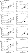
M2Rs and M4Rs are required for mAChR control of DA release in dorsal CPu. a–d, Mean peak [DA]o ± SEM versus frequency in CPu during four pulse trains (1–100 Hz) in control conditions (filled circles) or Oxo-M (unfilled) or Oxo-M plus DHβE (gray fill) normalized to [DA]o released by a single pulse in control conditions (left) or own condition (right), in wild types (WT) (a), M5R-knock-outs (M5R KO) (b), M2R-knock-outs (M2R KO) (c), and M4R-knock-outs (M4R KO) (d) (n = 9–19). The asterisks indicate significance level in post hoc Bonferroni's t tests for Oxo-M versus controls: **p < 0.01, ***p < 0.001. The daggers indicate significance level in Bonferroni's post hoc t tests for Oxo-M versus Oxo-M plus DHβE: ††p < 0.01, †††p < 0.001. Significance levels for comparison of Oxo-M plus DHβE versus control, or for data in right-hand panels, are omitted for clarity. a, b, In wild types and M5R-nulls, Oxo-M significantly modifies evoked [DA]o in an activity-dependent manner, whereas additional application of DHβE only slightly modifies evoked [DA]o further (n = 9). c, d, In M2R-KO or M4R-KO, Oxo-M does not significantly modify evoked [DA]o, but subsequent addition of DHβE does modify [DA]o in a marked and significant, activity-dependent manner (n = 15–19).
Figure 6.
Figure 6.
M4Rs (and not M2Rs) are required for mAChR control of DA release in NAc core. a–d, Mean peak [DA]o ± SEM versus frequency in NAc core during four pulse trains (1–100 Hz) in control conditions (filled circles) or Oxo-M (unfilled) or Oxo-M plus DHβE (gray fill) normalized to [DA]o released by a single pulse in control conditions (left) or own condition (right), in wild types (WT) (a), M5R-knock-outs (M5R KO) (b), M2R-knock-outs (M2R KO) (c), and M4R-knock-outs (M4R KO) (d) (n = 9–12). The asterisks indicate significance level in post hoc t tests for Oxo-M versus controls: *p < 0.05, ***p < 0.001. The daggers indicate significance level in post hoc t tests for Oxo-M versus Oxo-M plus DHβE: †††p < 0.001. Significance levels for comparison of Oxo-M plus DHβE versus control, or for data in right-hand panels, are omitted for clarity. a–c, In wild types, M5R-KO, or M2R-KO, Oxo-M significantly modifies evoked [DA]o in an activity-dependent manner and additional application of DHβE does not modify evoked [DA]o further (n = 9–12). d, In M4R-KO, Oxo-M does not modify evoked [DA]o, but subsequent addition of DHβE modifies [DA]o in a marked and significant, activity-dependent manner (n = 12).
Figure 7.
Figure 7.
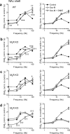
M4Rs (and not M2Rs) are required for mAChR control of DA release in NAc shell. a–d, Mean peak [DA]o ± SEM versus frequency in NAc shell during four pulse trains (1–100 Hz) in control conditions (filled circles) or Oxo-M (unfilled) or Oxo-M plus DHβE (gray fill) normalized to [DA]o released by a single pulse in control conditions (left) or own condition (right), in wild types (WT) (a), M5R-knock-outs (M5R KO) (b), M2R-knock-outs (M2R KO) (c), and M4R-knock-outs (M4R KO) (d) (n = 9). The asterisks indicate significance level in post hoc t tests for Oxo-M versus controls: *p < 0.05, **p < 0.01, ***p < 0.001. The daggers indicate significance level in post hoc t tests for Oxo-M versus Oxo-M plus DHβE: p < 0.05, ††p < 0.01, †††p < 0.001. Significance levels for comparison of Oxo-M plus DHβE versus control, or for data in right-hand panels, are omitted for clarity. a–c, In wild types, M5R-KO, or M2R-KO, Oxo-M significantly modifies evoked [DA]o in an activity-dependent manner, and additional application of DHβE only slightly modifies evoked [DA]o further (n = 9). d, In M4R-KO, Oxo-M does not modify evoked [DA]o, but subsequent addition of DHβE modifies [DA]o in a marked and significant, activity-dependent manner (n = 9).

Similar articles

Cited by

References

    1. Abeliovich A, Schmitz Y, Fariñas I, Choi-Lundberg D, Ho WH, Castillo PE, Shinsky N, Verdugo JM, Armanini M, Ryan A, Hynes M, Phillips H, Sulzer D, Rosenthal A. Mice lacking alpha-synuclein display functional deficits in the nigrostriatal dopamine system. Neuron. 2000;25:239–252. - PubMed
    1. Alcantara AA, Mrzljak L, Jakab RL, Levey AI, Hersch SM, Goldman-Rakic PS. Muscarinic m1 and m2 receptor proteins in local circuit and projection neurons of the primate striatum: anatomical evidence for cholinergic modulation of glutamatergic prefronto-striatal pathways. J Comp Neurol. 2001;434:445–460. - PubMed
    1. Aosaki T, Tsubokawa H, Ishida A, Watanabe K, Graybiel AM, Kimura M. Responses of tonically active neurons in the primate's striatum undergo systematic changes during behavioral sensorimotor conditioning. J Neurosci. 1994;14:3969–3984. - PMC - PubMed
    1. Apicella P. Tonically active neurons in the primate striatum and their role in the processing of information about motivationally relevant events. Eur J Neurosci. 2002;16:2017–2026. - PubMed
    1. Avshalumov MV, Chen BT, Marshall SP, Peña DM, Rice ME. Glutamate-dependent inhibition of dopamine release in striatum is mediated by a new diffusible messenger, H2O2. J Neurosci. 2003;23:2744–2750. - PMC - PubMed

Publication types

MeSH terms

LinkOut - more resources