

Internal and external features of the face are represented holistically in face-selective regions of visual cortex
- PMID: 20203214
- PMCID: PMC2839485
- DOI: 10.1523/JNEUROSCI.4863-09.2010
Internal and external features of the face are represented holistically in face-selective regions of visual cortex
Abstract
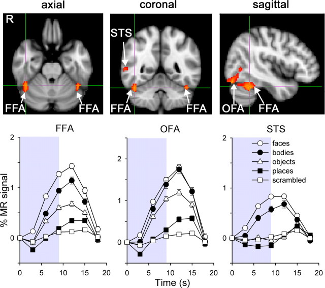
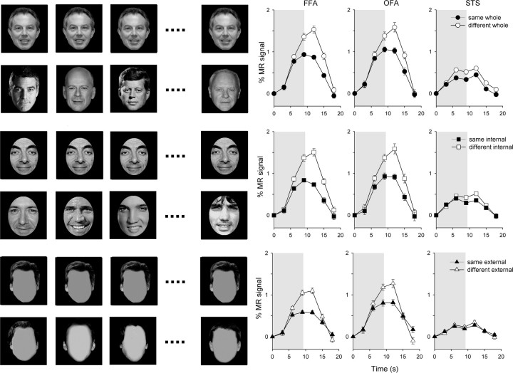
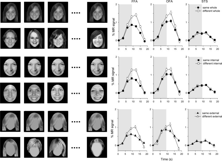
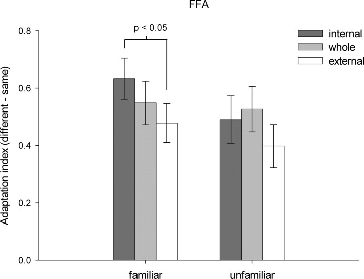
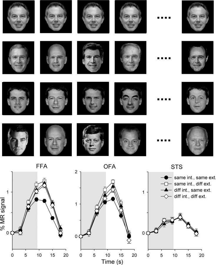
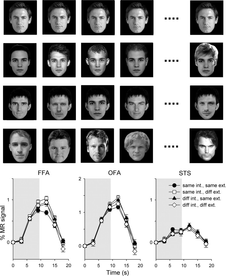
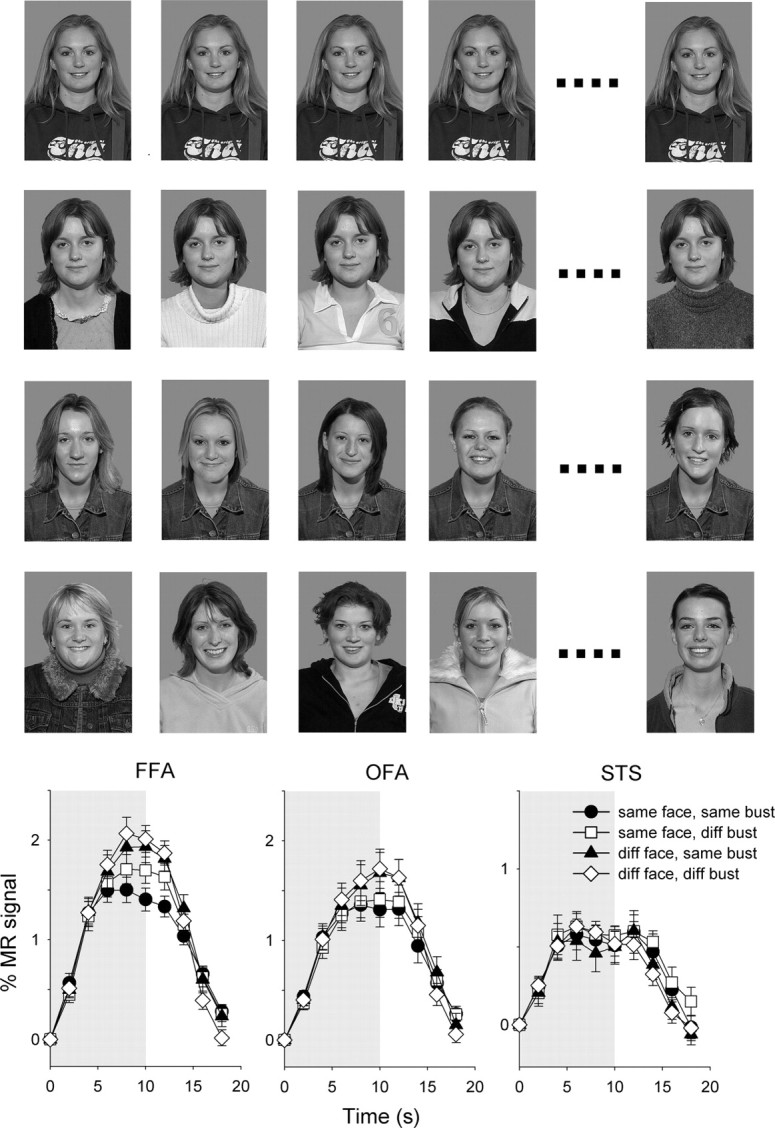
The perception and recognition of familiar faces depends critically on an analysis of the internal features of the face (eyes, nose, mouth). We therefore contrasted how information about the internal and external (hair, chin, face outline) features of familiar and unfamiliar faces is represented in face-selective regions. There was a significant response to both the internal and external features of the face when presented in isolation. However, the response to the internal features was greater than the response to the external features. There was significant adaptation to repeated images of either the internal or external features of the face in the fusiform face area (FFA). However, the magnitude of this adaptation was greater for the internal features of familiar faces. Next, we asked whether the internal features of the face are represented independently from the external features. There was a release from adaptation in the FFA to composite images in which the internal features were varied but the external features were unchanged, or when the internal features were unchanged but the external features varied, demonstrating a holistic response. Finally, we asked whether the holistic response to faces could be influenced by the context in which the face was presented. We found that adaptation was still evident to composite images in which the face was unchanged but body features were varied. Together, these findings show that although internal features are important in the neural representation of familiar faces, the face's internal and external features are represented holistically in face-selective regions of the human brain.
Figures
Comment in
-
The fusiform face area: in quest of holistic face processing.J Neurosci. 2010 Jun 30;30(26):8699-701. doi: 10.1523/JNEUROSCI.1921-10.2010. J Neurosci. 2010. PMID: 20592190 Free PMC article. No abstract available.
References
-
- Andrews TJ, Ewbank MP. Distinct representations for facial identity and changeable aspects of faces in the human temporal lobe. Neuroimage. 2004;23:905–913. - PubMed
-
- Bruce V, Young A. Understanding face recognition. Br J Psychol. 1986;77:305–327. - PubMed
-
- Burton AM, Jenkins R, Hancock PJB, White D. Robust representations for face recognition: the power of averages. Cogn Psychol. 2005;51:256–284. - PubMed
-
- Cox D, Meyers E, Sinha P. Contextually evoked object-specific responses in human visual cortex. Science. 2004;304:115–117. - PubMed
Publication types
MeSH terms
Grants and funding
LinkOut - more resources
Full Text Sources
Molecular Biology Databases