

Pim-1 kinase protects mitochondrial integrity in cardiomyocytes
- PMID: 20203306
- PMCID: PMC2864233
- DOI: 10.1161/CIRCRESAHA.109.212035
Pim-1 kinase protects mitochondrial integrity in cardiomyocytes
Abstract
Rationale: Cardioprotective signaling mediates antiapoptotic actions through multiple mechanisms including maintenance of mitochondrial integrity. Pim-1 kinase is an essential downstream effector of AKT-mediated cardioprotection but the mechanistic basis for maintenance of mitochondrial integrity by Pim-1 remains unexplored. This study details antiapoptotic actions responsible for enhanced cell survival in cardiomyocytes with elevated Pim-1 activity.
Objective: The purpose of this study is to demonstrate that the cardioprotective kinase Pim-1 acts to inhibit cell death by preserving mitochondrial integrity in cardiomyocytes.
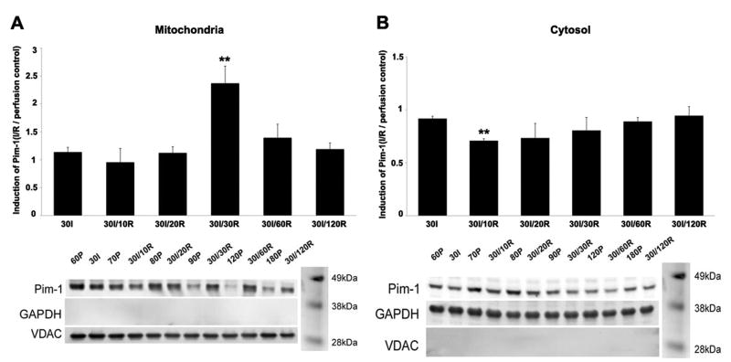
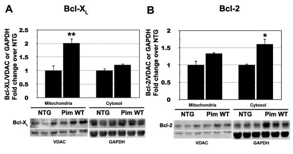
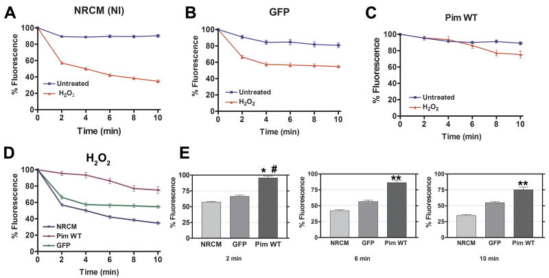
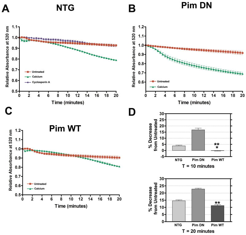
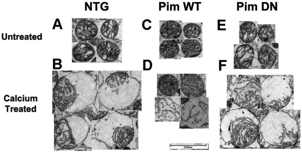
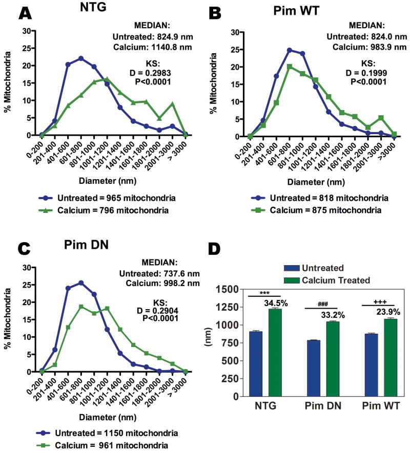
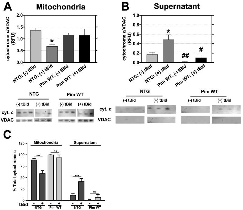
Methods and results: A combination of biochemical, molecular, and microscopic analyses demonstrate beneficial effects of Pim-1 on mitochondrial integrity. Pim-1 protein level increases in the mitochondrial fraction with a corresponding decrease in the cytosolic fraction of myocardial lysates from hearts subjected to 30 minutes of ischemia followed by 30 minutes of reperfusion. Cardiac-specific overexpression of Pim-1 results in higher levels of antiapoptotic Bcl-X(L) and Bcl-2 compared to samples from normal hearts. In response to oxidative stress challenge, Pim-1 preserves the inner mitochondrial membrane potential. Ultrastructure of the mitochondria is maintained by Pim-1 activity, which prevents swelling induced by calcium overload. Finally, mitochondria isolated from hearts created with cardiac-specific overexpression of Pim-1 show inhibition of cytochrome c release triggered by a truncated form of proapoptotic Bid.
Conclusion: Cardioprotective action of Pim-1 kinase includes preservation of mitochondrial integrity during cardiomyopathic challenge conditions, thereby raising the potential for Pim-1 kinase activation as a therapeutic interventional approach to inhibit cell death by antagonizing proapoptotic Bcl-2 family members that regulate the intrinsic apoptotic pathway.
Conflict of interest statement
Figures
Similar articles
-
Kaempferol protects cardiomyocytes against anoxia/reoxygenation injury via mitochondrial pathway mediated by SIRT1.Eur J Pharmacol. 2015 Aug 15;761:245-53. doi: 10.1016/j.ejphar.2015.05.056. Epub 2015 Jun 15. Eur J Pharmacol. 2015. PMID: 26086862
-
Postconditioning induces an anti-apoptotic effect and preserves mitochondrial integrity in isolated rat hearts.Biochim Biophys Acta. 2009 Jul;1787(7):794-801. doi: 10.1016/j.bbabio.2009.03.013. Epub 2009 Mar 26. Biochim Biophys Acta. 2009. PMID: 19328770
-
Glucosamine protects neonatal cardiomyocytes from ischemia-reperfusion injury via increased protein O-GlcNAc and increased mitochondrial Bcl-2.Am J Physiol Cell Physiol. 2008 Jun;294(6):C1509-20. doi: 10.1152/ajpcell.00456.2007. Epub 2008 Mar 26. Am J Physiol Cell Physiol. 2008. PMID: 18367586 Free PMC article.
-
Mitochondrial integrity: preservation through Akt/Pim-1 kinase signaling in the cardiomyocyte.Expert Rev Cardiovasc Ther. 2009 Aug;7(8):929-38. doi: 10.1586/erc.09.48. Expert Rev Cardiovasc Ther. 2009. PMID: 19673671 Free PMC article. Review.
-
Akt mediated mitochondrial protection in the heart: metabolic and survival pathways to the rescue.J Bioenerg Biomembr. 2009 Apr;41(2):169-80. doi: 10.1007/s10863-009-9205-y. J Bioenerg Biomembr. 2009. PMID: 19377835 Free PMC article. Review.
Cited by
-
PIM1 Promotes Survival of Cardiomyocytes by Upregulating c-Kit Protein Expression.Cells. 2020 Aug 31;9(9):2001. doi: 10.3390/cells9092001. Cells. 2020. PMID: 32878131 Free PMC article.
-
Enhancing the potential of cardiac progenitor cells: pushing forward with Pim-1.Circ Res. 2012 Apr 27;110(9):1154-6. doi: 10.1161/CIRCRESAHA.112.269183. Circ Res. 2012. PMID: 22539751 Free PMC article. No abstract available.
-
Salicylic diamines selectively eliminate residual undifferentiated cells from pluripotent stem cell-derived cardiomyocyte preparations.Sci Rep. 2021 Jan 27;11(1):2391. doi: 10.1038/s41598-021-81351-z. Sci Rep. 2021. PMID: 33504837 Free PMC article.
-
Pim-1 preserves mitochondrial morphology by inhibiting dynamin-related protein 1 translocation.Proc Natl Acad Sci U S A. 2013 Apr 9;110(15):5969-74. doi: 10.1073/pnas.1213294110. Epub 2013 Mar 25. Proc Natl Acad Sci U S A. 2013. PMID: 23530233 Free PMC article.
-
Functional Effect of Pim1 Depends upon Intracellular Localization in Human Cardiac Progenitor Cells.J Biol Chem. 2015 May 29;290(22):13935-47. doi: 10.1074/jbc.M114.617431. Epub 2015 Apr 16. J Biol Chem. 2015. PMID: 25882843 Free PMC article.
References
-
- Rosamond W, Flegal K, Friday G, Furie K, Go A, Greenlund K, Haase N, Ho M, Howard V, Kissela B, Kittner S, Lloyd-Jones D, Mc Dermott M, Meigs J, Moy C, Nichol G, O'Donnell CJ, Roger V, Rumsfeld J, Sorlie P, Steinberger J, Thom T, Wasserthiel-Smoller S, Hong Y. Heart disease and stroke statistics—2007 update: a Statistics Subcommitte. Circulation. 2007;115:e69–171. - PubMed
-
- Narula J, Pandey P, Arbustini E, Haider N, Narula N, Kolodgie FD, Dal Bello B, Semigran MJ, Bielsa-Masdeu A, Dec GW, Israels S, Ballester M, Virmani R, Saxena S, Kharbanda S. Apoptosis in heart failure: release of cytochrome c from mitochondria and activation of caspase-3 in human cardiomyopathy. Proc Natl Acad Sci USA. 1999;96:8144–8149. - PMC - PubMed
-
- Steenbergen C, Afshari CA, Petranka JG, Collins J, Martin K, Bennett L, Haugen A, Bushel P, Murphy E. Alterations in apoptotic signaling in human idiopathic cardiomyopathic hearts in failure. Am J Physiol Heart Circ Physiol. 2003;284:H268–H276. - PubMed
-
- Olivetti G, Quaini F, Sala R, Lagrasta C, Corradi D, Bonacina E, Gambert SR, Cigola E, Anversa P. Acute myocardial infarction in humans is associated with activation of programmed myocyte cell death in the surviving portion of the heart. J Mol Cell Cardiol. 1996;28:2005–2016. - PubMed
Publication types
MeSH terms
Substances
Grants and funding
- 1R37HL091102/HL/NHLBI NIH HHS/United States
- RC1 HL100891/HL/NHLBI NIH HHS/United States
- R01 HL075573/HL/NHLBI NIH HHS/United States
- R01 HL060590/HL/NHLBI NIH HHS/United States
- 1R01HL091102/HL/NHLBI NIH HHS/United States
- 1P01AG023071/AG/NIA NIH HHS/United States
- R21 HL102714/HL/NHLBI NIH HHS/United States
- 5R01HL067245/HL/NHLBI NIH HHS/United States
- 1P01HL085577/HL/NHLBI NIH HHS/United States
- P01 AG023071/AG/NIA NIH HHS/United States
- R37 HL091102/HL/NHLBI NIH HHS/United States
- R01 HL104535/HL/NHLBI NIH HHS/United States
- R01 HL067245/HL/NHLBI NIH HHS/United States
- P01 HL085577/HL/NHLBI NIH HHS/United States
- R21 HL104544/HL/NHLBI NIH HHS/United States
- R01 AG033283/AG/NIA NIH HHS/United States
LinkOut - more resources
Full Text Sources
Molecular Biology Databases
Research Materials
