Review
. 2010 Mar 5;106(4):633-46.
doi: 10.1161/CIRCRESAHA.109.207381.

S-nitrosylation in cardiovascular signaling

Affiliations
Review

S-nitrosylation in cardiovascular signaling

Brian Lima et al. Circ Res. .

Abstract

Well over 2 decades have passed since the endothelium-derived relaxation factor was reported to be the gaseous molecule nitric oxide (NO). Although soluble guanylyl cyclase (which generates cyclic guanosine monophosphate, cGMP) was the first identified receptor for NO, it has become increasingly clear that NO exerts a ubiquitous influence in a cGMP-independent manner. In particular, many, if not most, effects of NO are mediated by S-nitrosylation, the covalent modification of a protein cysteine thiol by an NO group to generate an S-nitrosothiol (SNO). Moreover, within the current framework of NO biology, endothelium-derived relaxation factor activity (ie, G protein-coupled receptor-mediated, or shear-induced endothelium-derived NO bioactivity) is understood to involve a central role for SNOs, acting both as second messengers and signal effectors. Furthermore, essential roles for S-nitrosylation have been implicated in virtually all major functions of NO in the cardiovascular system. Here, we review the basic biochemistry of S-nitrosylation (and denitrosylation), discuss the role of S-nitrosylation in the vascular and cardiac functions of NO, and identify current and potential clinical applications.


Figures

Figure 1
Figure 1
The roles of cGMP and S-nitrosylation in NO-based signaling (A) and enzymatic protein denitrosylation mediated by the S-nitrosoglutathione reductase (GSNOR) and thioredoxin (Trx) systems (B). (A) NO synthase (NOS) synthesizes NO, which may activate soluble guanylyl cyclase and thereby enhance production of cGMP (left) or subserve protein S-nitrosylation (right). The cGMP-dependent pathway is deactivated by cGMP-phosphodiesterase (PDE), which hydrolyzes cGMP to GMP (PDE may also be activated allosterically by cGMP). The SNO-based mechanisms are dynamically regulated via S-nitrosylation and denitrosylation of a multitude of cysteine-containing proteins. In contrast to the multiple elements regulated by S-nitrosylation, the cGMP-based signaling system relies primarily on the cGMP-dependent protein kinase, PKG. (B) Proteins undergo reversible S-nitrosylation and denitrosylation (center). Denitrosylation mediated by GSNOR is depicted on the left. Transnitrosylation of glutathione (GSH) by S-nitrosylated proteins generates GSNO and native protein. GSNO undergoes NADH-dependent reduction by GSNOR to generate glutathione S-hydroxysulfenamide (GSNHOH), which can undergo further reaction with GSH to generate oxidized glutathione (GSSG). The redox cycle is completed by reduction of GSSG to GSH via GSSG reductase. Denitrosylation mediated by the thioredoxin (Trx) system is depicted on the right. The active site dithiol motif (CXXC) of Trx1 (cytoplasmic) or Trx2 (mitochondrial) undergoes oxidation coupled to denitrosylation of SNO substrate. Oxidized Trx is reduced by the selenoprotein thioredoxin reductase (TrxR), which employs the reducing power of NADPH to regenerate active Trx.
Figure 2
Figure 2
S-nitrosylation of NSF is anti-inflammatory and anti-thrombotic, . In endothelial cells, inhibitory S-nitrosylation of NSF suppresses exocytosis of Weibel-Palade bodies and thereby externalization of P-selectin, which inhibits leukocyte rolling and thus vascular inflammation. Similarly, in platelets (labeling in parentheses), inhibitory S-nitrosylation of NSF suppresses exocytosis of secretory granules and thereby externalization of P-selectin (and other adhesive molecules), which reduce platelet activation, adhesion, aggregation and rolling on the endothelium. These effects are anti-thrombotic.
Figure 3
Figure 3
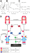
SNO-Hb subserves hypoxic vasodilation. (A) PO2 determines the ability of RBCs to constrict or relax aortic ring preparations on a second-by-second time scale. PO2 is indicated for each curve, which illustrate a graded response. (B) and (C) O2-dependent effects of SNO-Hb and Hb on local cerebral blood flow are shown in normoxia and hyperoxia. SNO-Hb infusion in vivo (1µmol/kg over 3min, beginning at time 0) immediately increases local cerebral blood flow in the caudate-putamen nucleus of rats breathing 21% O2 at 1 atmosphere absolute (ATA), where tissue PO2 ranges from 19 to 37 mmHg. Thus, SNO-Hb appropriately increases blood flow in relatively hypoxic tissue; however, non-nitrosylated Hb decreases perfusion. In 100% O2 at 3 ATA, where tissue PO2 ranges from 365 to 538 mmHg, vasodilation is abrogated because SNO-Hb cannot allosterically dispense NO bioactivity (Adapted from Allen et al.). (D) Allosteric transitions of circulating hemoglobin (Hb) regulate delivery of NO bioactivity to preserve vascular O2 homeostasis. Hb in RBCs senses [O2] and responds through allosterically controlled NO binding, SNO formation, and NO group release. At high O2 in the pulmonary venous system, Hb is in the R-state, Cys β93 is reactive and Cys93-SNO is shielded in a hydrophobic pocket. On partial RBC deoxygenation in the periphery, Hb adopts the T configuration and Cys β93-SNO is exposed to solvent. Further, in venous blood a population of deoxygenated (T-state) Hb reacts with NO to produce nitrosylated heme in the β-chain (bottom left). Transition to R-state draws Cys β93 close to the nitrosylated heme (top left) with a subsequent transfer of NO from heme to Cys β93, forming a SNO (top right). Deoxygenation of Hb favors the T conformation (bottom right), allowing SNO-Cys β93 to react with other cellular thiols, and thereby facilitating release of NO/SNO from the RBC. (Adapted from 126).
Figure 4
Figure 4
S-nitrosylation regulates myocardial Ca2+ handling and thereby excitation-contraction coupling. RyR2, the cardiac form of the tetrameric ryanodine receptor/Ca2+ release channel, is localized to the SR membrane in proximity with the plasma membrane L-type calcium channel (LTCC), which provides the substrate for calcium-mediated calcium release from SR to cytosol. The SR-localized Ca2+-ATPase (SERCA) replenishes SR Ca2+. RyR2 co-localizes with nNOS in the SR, and S-nitrosylation of RyR2 (mediated by GSNO) potentiates Ca2+ release. As in skeletal muscle RyR1, physiological S-nitrosylation of one or a few Cys within each RyR2 monomer is likely to be the case. S-nitrosylation of the LTCC (α1C subunit; resulting in, for example, attenuated β-AR-dependent contractility) and of SERCA is inhibitory. Hypo-S-nitrosylation of RyR2 is associated with diastolic Ca2+ leakage and arrhythmia characteristic of sudden cardiac death. S-nitrosylation of the LTCC has been associated with ischemic preconditioning that reduces reperfusion injury, whereas hyper-S-nitrosylation of the LTCC has been associated with atrial fibrillation. Note in addition that aberrant S-nitrosylation can result from the translocation of nNOS to the plasma membrane that is seen in association with myocardial infarction and cardiomyopathy. Further, aberrant and in particular hyper-S-nitrosylation can result in irreversible oxidative modification of S-nitrosylated proteins in concert with reactive oxygen species produced by endogenous enzymes including xanthine oxidase.
Figure 5
Figure 5
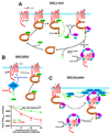
A Schematic summary of the regulation of agonist-induced β2-adrenergic receptor trafficking by S-nitrosylation/denitrosylation of β-arrestin 2 (β-Arr 2), G protein-coupled receptor kinase 2 (GRK2), and dynamin. (A) β-Arr 2 serves as a scaffold that functionally colocalizes eNOS and β-ARs (as well as other G protein-coupled receptors [GPCRs]). Ligand (isoproterenol) stimulation results in activation of eNOS and S-nitrosylation of β-Arr 2. S-nitrosylation of β-Arr 2 promotes its dissociation from eNOS and its association with clathrin heavy chain/β-adaptin, which facilitates routing of the β2-AR into the clathrin-based endocytotic pathway, and β-Arr 2 is subsequently denitrosylated. (B) Inhibition of GRK2 by ligand-coupled S-nitrosylation suppresses agonist-stimulated β-AR phosphorylation, β-Arr 2 recruitment, and receptor desensitization and downregulation (schematic at top) At bottom: desensitization (decline in cardiac contractility in the continued presence of ISO) is enhanced by inhibiting NO production. (C) After GPCR activation, eNOS-mediated S-nitrosylation of dynamin promotes multimerization and GTPase activity, as well as relocation to the plasma membrane, which facilitates scission of endocytotic vesicles and receptor internalization. (Adapted from28, 29).
Figure 6
Figure 6
S-nitrosylation of channel proteins regulates all phases of the ventricular action potential. S-nitrosylation of SCN5A channels enhances the Na+ current (INa), , whereas S-nitrosylation of the α1C subunit of the L-type Ca2+ channel inhibits the L-type Ca2+ current (ICaL), . Among voltage-gated potassium channels, S-nitrosylation of the KCNQ1 subunit facilitates the slowly activating component of the delayed rectifier K+ current (IKs), whereas S-nitrosylation exerts an inhibitory influence on Kv4.3 and thus the transient outward potassium current (ITo), as well as Kir2.1, and thus IK1 (phase 4). In addition, heterologously expressed human ether-a-go-go-related gene 1 (hERG1) potassium channels, which mediate the rapidly activating delayed rectifier K+ channel (IKr) in their native environment, are inhibited by NO in a cGMP-independent fashion. Note that, in the atrium, S-nitrosylation inhibits hKv1.5 and thus the ultra-rapid delayed rectifier current (Adapted from138).

Comment in

Similar articles

Cited by

References

    1. Guyton AC, Coleman TG, Granger HJ. Circulation: overall regulation. Annu Rev Physiol. 1972;34:13–46. - PubMed
    1. McMahon TJ, Moon RE, Luschinger BP, Carraway MS, Stone AE, Stolp BW, Gow AJ, Pawloski JR, Watke P, Singel DJ, Piantadosi CA, Stamler JS. Nitric oxide in the human respiratory cycle. Nat Med. 2002;8:711–717. - PubMed
    1. Dzik S. Nitric oxide: nature's third respiratory gas. Transfusion. 2002;42:1532–1533. - PubMed
    1. Allen BW, Stamler JS, Piantadosi CA. Hemoglobin, nitric oxide and molecular mechanisms of hypoxic vasodilation. Trends Mol Med. 2009;15:452–460. - PMC - PubMed
    1. Bredt DS, Snyder SH. Nitric oxide: a physiologic messenger molecule. Annu Rev Biochem. 1994;63:175–195. - PubMed

Publication types

MeSH terms