Review
. 2010 Mar 5;106(4):674-85.
doi: 10.1161/CIRCRESAHA.109.212936.

Biological therapies for cardiac arrhythmias: can genes and cells replace drugs and devices?

Affiliations
Review

Biological therapies for cardiac arrhythmias: can genes and cells replace drugs and devices?

Hee Cheol Cho et al. Circ Res. .

Abstract

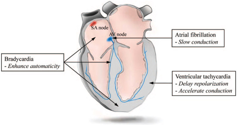
Cardiac rhythm disorders reflect failures of impulse generation and/or conduction. With the exception of ablation methods that yield selective endocardial destruction, present therapies are nonspecific and/or palliative. Progress in understanding the underlying biology opens up prospects for new alternatives. This article reviews the present state of the art in gene- and cell-based therapies to correct cardiac rhythm disturbances. We begin with the rationale for such approaches, briefly discuss efforts to address aspects of tachyarrhythmia, and review advances in creating a biological pacemaker to cure bradyarrhythmia. Insights gained bring the field closer to a paradigm shift away from devices and drugs, and toward biologics, in the treatment of rhythm disorders.


Conflict of interest statement

Disclosures

E.M. owns stock in Excigen, Inc which licensed intellectual property on biological pacemakers. No research funding was provided by Excigen. H.C.C. reports no conflict of interest.

Figures

Figure 1.
Figure 1.
Principles of biological approaches (in italics) recruited to treat major cardiac arrhythmias, and sites within the heart where the various strategies have been tested.
Figure 2.
Figure 2.
Suppression of Kir2.1 channels unmasks latent pacemaker activity in ventricular cells. A, APs evoked by depolarizing external stimuli in control ventricular myocytes. B, Spontaneous APs in Kir2.1AAA-transduced myocytes with depressed IK1. C, Baseline electrocardiograms in normal sinus rhythm. D, Ventricular rhythms 72 hours after gene transfer of Kir2.1AAA. P waves (A and arrow) and wide QRS complexes (V and arrow) march through to their own rhythm. A through D are reproduced from Miake et al with permission. E, Guinea pig ventricular myocytes in which Kir2.1AAA was overexpressed (left) and exposed acutely to 1 μmol/L isoproterenol (J. Miake, H. B. Nuss, and E.M., unpublished data, 2002).
Figure 3.
Figure 3.
Dynamics of induced reentrant spiral waves in 21-mm-diameter monolayers with a central island of Kir2.1 suppression. The mapping region is 17 mm in diameter. A, Island of Kir2.1-suppressed NRVMs (indicated by white circles) acting as a source of spontaneous activity, as seen by the spread of spontaneous cardiac impulses (shown by black arrows) from an interior region of the island. B, An induced spiral wave anchored momentarily to the region of IK1 suppression (shown by white circle). C, Transition of initial spiral wave into a multi-armed spiral wave. D, Transition of initial spiral wave into a 2-armed spiral wave. E, Transition of the 2-armed spiral wave of D into a figure-of-eight reentry that later drifted to the edge of the monolayer and terminated. Reproduced from Sekar et al with permission.
Figure 4.
Figure 4.
A, In vitro and in vivo fusion of myocytes with HCN1-fibroblasts. GFP-positive heterokaryons after in vitro (top) or in vivo (bottom) fusion of myocytes with HCN1-fibroblasts expressing GFP as a reporter. B, Heterokaryon formed by in vivo fusion of HCN1-fibroblast and a myocyte displays spontaneous AP oscillations in normal Tyrode’s (top). Presence of 1 mmol/L isoproterenol in the external solution increased the frequency of spontaneous AP oscillation in the same heterokaryon (bottom). C, ECGs from guinea pig hearts injected with HCN1-fibroblast cells. Top, Ectopic ventricular beats (diagonal arrows) are unleashed on slowing of the heart rate, which share the same polarity and morphology as the electrode-paced ECGs recorded at the site of HCN1-fibroblast injection. Bottom, In another animal, junctional escape rhythms (horizontal arrows) were overtaken by ectopic ventricular beats (diagonal arrows, 16 days after cell injection). Reproduced from Cho et al with permission.
Figure 5.
Figure 5.
Representative raw recordings from HEK293 cells transfected with the following constructs. A, Voltage-clamp recordings from HEK293 cells transfected with either NaChBac (left), HERG (middle), or Kir2.1 (right). Dotted line indicates zero current level. B, APs from 3 different cells during current-clamp recordings. Each cell expressed all 3 channels, NaChBac, HERG, and Kir2.1. Dotted line indicates zero mV potential. C, Spontaneous APs recorded from a representative cell transfected with single plasmid expressing HCN1, NaChBac, and Kir2.1. Adapted from Cho et al.
Figure 6.
Figure 6.
A summary of different approaches to creating a biological pacemaker. First approach (top row) is a strict gene therapy in which Kir2.1AAA, HCN, or synthetic pacemaker channel genes are overexpressed in myocytes via adenoviral delivery. Kir2.1 dominant negative proteins suppress repolarizing, outward currents whereas pacemaker channels directly contribute to diastolic membrane potential depolarization. Delivering If by MSCs requires gap-junctional coupling between myocytes and MSCs (second row). In the cell fusion approach (third row), If and the pacemaker activity arise from the HCN channels expressed on the cell membrane of the heterokaryon, without the need for gap-junctional coupling. Spontaneously beating human EBs and cardiospheres transduce their pacemaker activity to cardiomyocytes via electrotonic cell–cell coupling (fourth row).

Similar articles

Cited by

References

    1. Coplen SE, Antman EM, Berlin JA, Hewitt P, Chalmers TC. Efficacy and safety of quinidine therapy for maintenance of sinus rhythm after cardioversion. A meta-analysis of randomized control trials. Circulation. 1990;82:1106–1116. - PubMed
    1. Echt DS, Liebson PR, Mitchell LB, Peters RW, ObiasManno D, Barker AH, Arensberg D, Baker A, Friedman L, Green HL, Huther ML, Richardson DW. Mortality and morbidity in patients receiving encainide, flecainide, or placebo. The Cardiac Arrhythmia Suppression Trial. N Engl J Med. 1991;324:781–788. - PubMed
    1. Siebels J, Cappato R, Ruppel R, Schneider MA, Kuck KH. Preliminary results of the Cardiac Arrest Study Hamburg (CASH). CASH Investigators. Am J Cardiol. 1993;72:109F–113F. - PubMed
    1. Waldo AL, Camm AJ, deRuyter H, Friedman PL, MacNeil DJ, Pauls JF, Pitt B, Pratt CM, Schwartz PJ, Veltri EP. Effect of d-sotalol on mortality in patients with left ventricular dysfunction after recent and remote myocardial infarction. The SWORD Investigators. Survival With Oral d-Sotalol. Lancet. 1996;348:7–12. - PubMed
    1. Falk RH. Atrial fibrillation. N Engl J Med. 2001;344:1067–1078. - PubMed

Publication types

Substances