

Circulating mitochondrial DAMPs cause inflammatory responses to injury
- PMID: 20203610
- PMCID: PMC2843437
- DOI: 10.1038/nature08780
Circulating mitochondrial DAMPs cause inflammatory responses to injury
Abstract
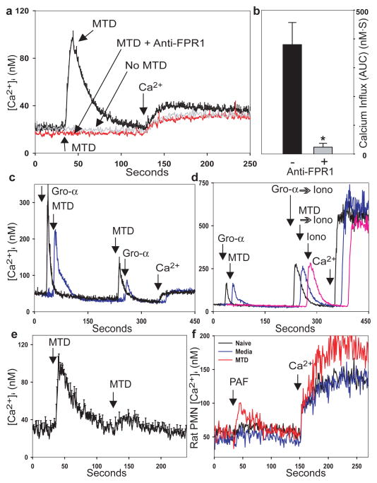
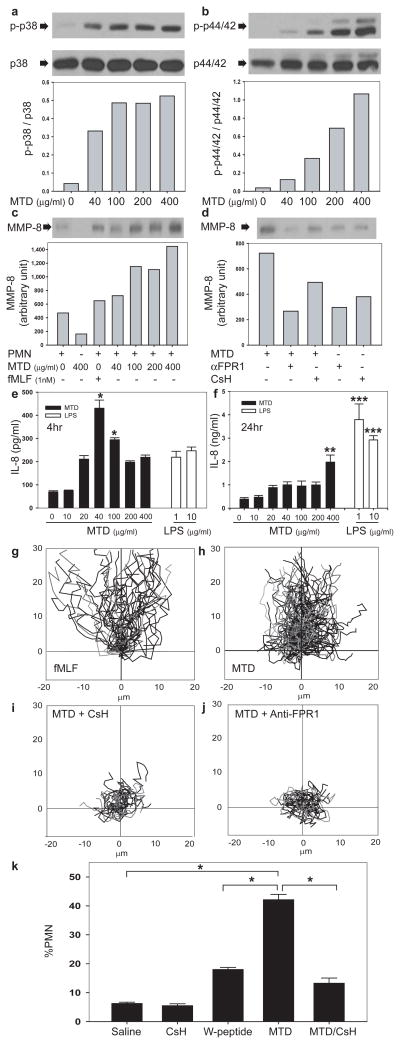
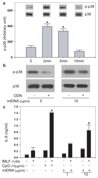
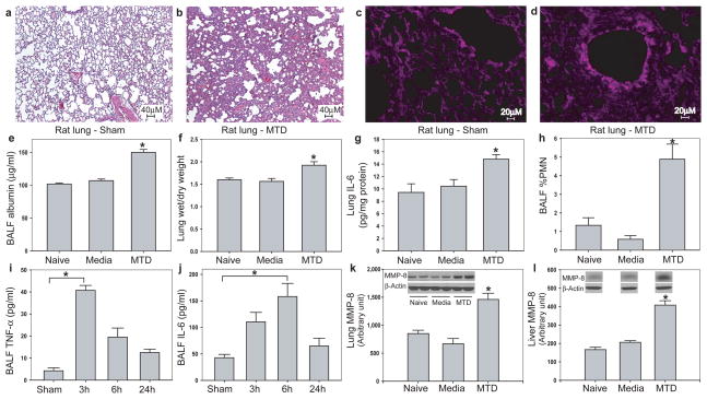
Injury causes a systemic inflammatory response syndrome (SIRS) that is clinically much like sepsis. Microbial pathogen-associated molecular patterns (PAMPs) activate innate immunocytes through pattern recognition receptors. Similarly, cellular injury can release endogenous 'damage'-associated molecular patterns (DAMPs) that activate innate immunity. Mitochondria are evolutionary endosymbionts that were derived from bacteria and so might bear bacterial molecular motifs. Here we show that injury releases mitochondrial DAMPs (MTDs) into the circulation with functionally important immune consequences. MTDs include formyl peptides and mitochondrial DNA. These activate human polymorphonuclear neutrophils (PMNs) through formyl peptide receptor-1 and Toll-like receptor (TLR) 9, respectively. MTDs promote PMN Ca(2+) flux and phosphorylation of mitogen-activated protein (MAP) kinases, thus leading to PMN migration and degranulation in vitro and in vivo. Circulating MTDs can elicit neutrophil-mediated organ injury. Cellular disruption by trauma releases mitochondrial DAMPs with evolutionarily conserved similarities to bacterial PAMPs into the circulation. These signal through innate immune pathways identical to those activated in sepsis to create a sepsis-like state. The release of such mitochondrial 'enemies within' by cellular injury is a key link between trauma, inflammation and SIRS.
Figures
Comment in
-
Clinical immunology: Culprits with evolutionary ties.Nature. 2010 Mar 4;464(7285):41-2. doi: 10.1038/464041a. Nature. 2010. PMID: 20203598 Free PMC article.
References
-
- Bone RC. Toward an epidemiology and natural history of SIRS (systemic inflammatory response syndrome) JAMA. 1992;268:3452–5. - PubMed
-
- Janeway CA., Jr Approaching the asymptote? Evolution and revolution in immunology. Cold Spring Harb Symp Quant Biol. 1989;54(Pt 1):1–13. - PubMed
-
- Matzinger P. Tolerance, danger, and the extended family. Annu Rev Immunol. 1994;12:991–1045. - PubMed
-
- Sagan L. On the origin of mitosing cells. J Theor Biol. 1967;14:255–74. - PubMed
-
- Sasser SM, et al. Guidelines for field triage of injured patients. Recommendations of the National Expert Panel on Field Triage. MMWR Recomm Rep. 2009;58:1–35. - PubMed
Publication types
MeSH terms
Substances
Grants and funding
LinkOut - more resources
Full Text Sources
Other Literature Sources
Medical
Molecular Biology Databases
Miscellaneous
