. 2010 Mar 4;464(7285):121-5.
doi: 10.1038/nature08778.

SIRT3 regulates mitochondrial fatty-acid oxidation by reversible enzyme deacetylation

Affiliations

SIRT3 regulates mitochondrial fatty-acid oxidation by reversible enzyme deacetylation

Matthew D Hirschey et al. Nature. .

Abstract

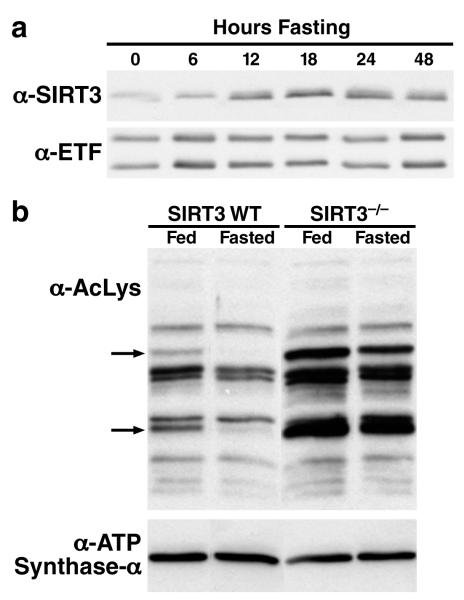
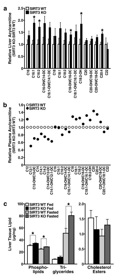
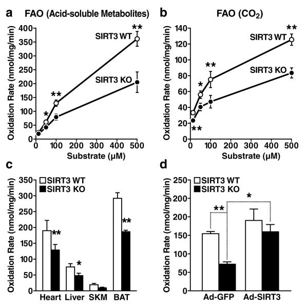
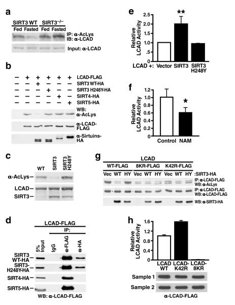
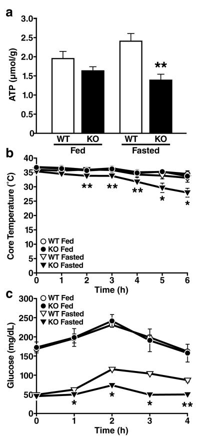
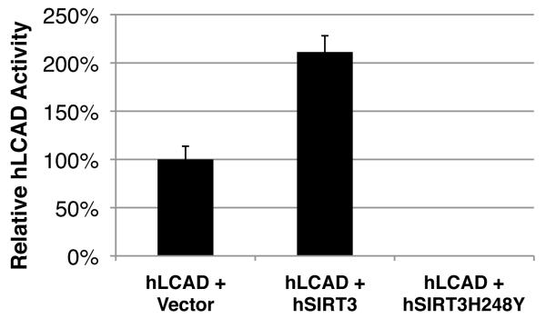
Sirtuins are NAD(+)-dependent protein deacetylases. They mediate adaptive responses to a variety of stresses, including calorie restriction and metabolic stress. Sirtuin 3 (SIRT3) is localized in the mitochondrial matrix, where it regulates the acetylation levels of metabolic enzymes, including acetyl coenzyme A synthetase 2 (refs 1, 2). Mice lacking both Sirt3 alleles appear phenotypically normal under basal conditions, but show marked hyperacetylation of several mitochondrial proteins. Here we report that SIRT3 expression is upregulated during fasting in liver and brown adipose tissues. During fasting, livers from mice lacking SIRT3 had higher levels of fatty-acid oxidation intermediate products and triglycerides, associated with decreased levels of fatty-acid oxidation, compared to livers from wild-type mice. Mass spectrometry of mitochondrial proteins shows that long-chain acyl coenzyme A dehydrogenase (LCAD) is hyperacetylated at lysine 42 in the absence of SIRT3. LCAD is deacetylated in wild-type mice under fasted conditions and by SIRT3 in vitro and in vivo; and hyperacetylation of LCAD reduces its enzymatic activity. Mice lacking SIRT3 exhibit hallmarks of fatty-acid oxidation disorders during fasting, including reduced ATP levels and intolerance to cold exposure. These findings identify acetylation as a novel regulatory mechanism for mitochondrial fatty-acid oxidation and demonstrate that SIRT3 modulates mitochondrial intermediary metabolism and fatty-acid use during fasting.


Figures

Figure 1
Figure 1. Fasting induces SIRT3 expression in oxidative tissues
a. Mitochondria were isolated from livers of fed or fasted (6-48 h) wt mice and analyzed for SIRT3 expression by western blot analysis, electron transfer flavoprotein (ETF) was used as a reference; b. Mitochondria isolated from livers of fed or fasted wt and SIRT3-/- mice were analyzed by western blotting analysis with an antiserum specific for anti-acetyllysine, ATP synthase alpha was used as a reference. The arrows identify candidate SIRT3 target proteins: deacetylated during fasting in wt mice, relatively hyperacetylated and not deacetylated during fasting in SIRT3-/- mice.
Figure 2
Figure 2. Abnormal accumulation of acylcarnitines and triglyceride in the livers of mice lacking SIRT3 during fasting
a, b. Metabolomic analyses were conducted on mouse liver tissue (a) and plasma (b), data obtained in SIRT3-/- mice are shown relative to wt mice (n=5/genotype, fasted 24 h); c. Livers extracts from SIRT3-/- and wt mice were analyzed for total phospholipids, triglycerides and cholesterol esters (n=5/genotype, fed or fasted 24 h), *p<0.05
Figure 3
Figure 3. Defective fatty acid oxidation in mice lacking SIRT3-/-
a, b. Fatty acid oxidation was measured by incubation of liver extract from wt and SIRT3-/- mice with 14C-palmitate, acid-soluble metabolites [(ASM), panel a] and captured CO2 (panel b), n=10/genotype; c. Mitochondrial fatty acid oxidation was measured in other oxidizing tissues, including heart, liver, mixed gastrocnemius and soleus skeletal muscle (SKM), and brown adipose tissue (BAT) (CO2, n=7/genotype, 100 μM substrate), d. Fatty acid oxidation was measured by incubation of 14C-palmitate (100 μM) in liver extract from wt and SIRT3-/- mice one week after injection with adenovirus expression vectors for GFP or SIRT3 (ASM, n=3-4/category); *p<0.05, **p<0.01.
Figure 4
Figure 4. LCAD is hyperacetylated in SIRT3-/-mice, deacetylated by SIRT3 in vivo and in vitro, and displays increased enzymatic activity when deacetylated
a. Liver extracts from wt and SIRT3-/- mice (fed or fasted) were immunoprecipitated with an anti-acetyllysine antiserum and analyzed with anti-LCAD antiserum; b. Expression vectors for wt SIRT3, SIRT3-H248Y (catalytically-inactive SIRT3 mutant), SIRT4, or SIRT5 were co-transfected with expression vectors for FLAG-tagged LCAD and the level of LCAD acetylation was assessed; c. Recombinant LCAD expressed in E. coli was incubated in vitro with recombinant SIRT3 or SIRT3-H248Y, and LCAD acetylation status was assessed; d. Expression vectors for wt SIRT3, SIRT3-H248Y, SIRT4 and SIRT5 (HA-tagged) were co-transfected with expression vectors for FLAG-tagged LCAD and assessed for interaction by co-immunoprecipitation; e. LCAD was expressed and purified with SIRT3 or SIRT3-H248Y and its enzymatic activity measured in vitro using 2, 6 dimethylheptanoyl-CoA as a substrate (n=4 independent assays); f. Recombinant LCAD was expressed in E. coli in the absence (Control) or presence of nicotinamide (NAM, 50 mM), purified and its enzymatic activity measured in vitro using 2, 6 dimethylheptanoyl-CoA as a substrate (n=4 independent assays); g. Expression vectors for wt LCAD, LCAD single acetylation point mutant (LCAD-K42R), or LCAD eight acetylation point mutant (LCAD-8KR) were co-transfected with expression vectors for wt SIRT3 or SIRT3-H248Y, and the level of acetylation was assessed; h. Wild-type LCAD, LCAD-K42R, or LCAD-8KR were expressed, and measured for enzymatic activity in vitro using 2, 6 dimethylheptanoyl-CoA as a substrate (n=5 measurement/sample, error bars represent data two independent protein purifications); *p<0.05, **p<0.01.
Figure 5
Figure 5. Mice lacking SIRT3 show reduced ATP production, cold intolerance and hypoglycemia
a. Hepatic ATP levels were measured in fed and fasted wt and SIRT3-/- mice (n=5/genotype/condition) b, c. Core temperature (b) and blood glucose (c) were measured in fed and fasted wt and SIRT3-/- mice exposed to cold (4°C) for 6 h (n=5/genotype/condition); *p<0.05, **p<0.01
Figure 6
Figure 6

Similar articles

Cited by

References

    1. Schwer B, Bunkenborg J, Verdin RO, Andersen JS, Verdin E. Reversible lysine acetylation controls the activity of the mitochondrial enzyme acetyl-CoA synthetase 2. Proc Natl Acad Sci U S A. 2006;103:10224–10229. - PMC - PubMed
    1. Hallows WC, Lee S, Denu JM. Sirtuins deacetylate and activate mammalian acetyl-CoA synthetases. Proc Natl Acad Sci U S A. 2006;103:10230–10235. - PMC - PubMed
    1. Lombard DB, et al. Mammalian Sir2 Homolog SIRT3 Regulates Global Mitochondrial Lysine Acetylation. Mol Cell Biol. 2007;27:8807–8814. - PMC - PubMed
    1. Kim SC, et al. Substrate and functional diversity of lysine acetylation revealed by a proteomics survey. Mol Cell. 2006;23:607–618. - PubMed
    1. Guarente L. Sirtuins as potential targets for metabolic syndrome. Nature. 2006;444:868–874. - PubMed

Publication types

MeSH terms