. 2010 Apr 7;29(7):1262-71.
doi: 10.1038/emboj.2010.23. Epub 2010 Mar 4.

The mycobacterial Mpa-proteasome unfolds and degrades pupylated substrates by engaging Pup's N-terminus

Affiliations

The mycobacterial Mpa-proteasome unfolds and degrades pupylated substrates by engaging Pup's N-terminus

Frank Striebel et al. EMBO J. .

Abstract

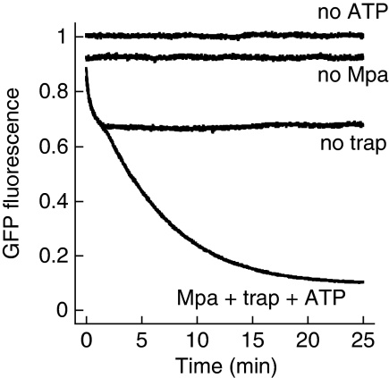
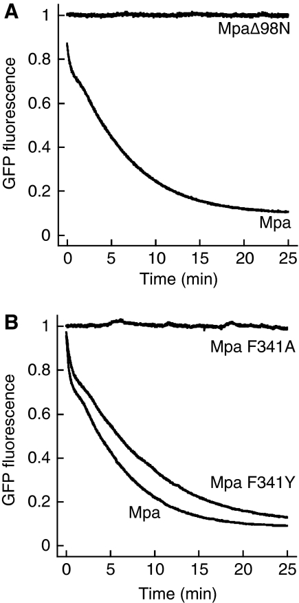
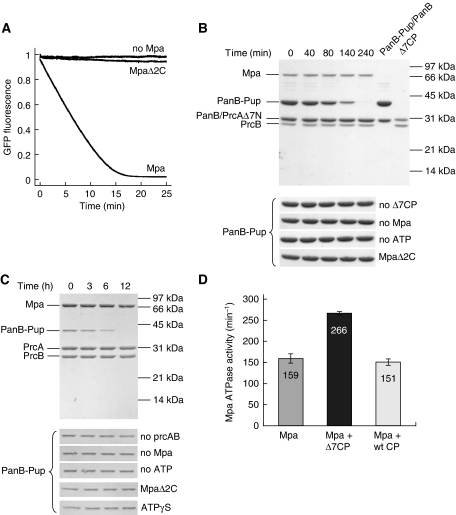
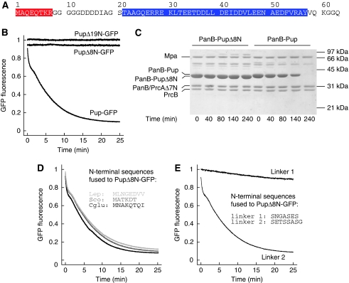
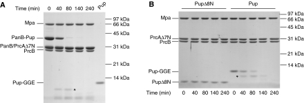
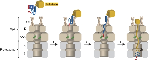
Mycobacterium tuberculosis, along with other actinobacteria, harbours proteasomes in addition to members of the general bacterial repertoire of degradation complexes. In analogy to ubiquitination in eukaryotes, substrates are tagged for proteasomal degradation with prokaryotic ubiquitin-like protein (Pup) that is recognized by the N-terminal coiled-coil domain of the ATPase Mpa (also called ARC). Here, we reconstitute the entire mycobacterial proteasome degradation system for pupylated substrates and establish its mechanistic features with respect to substrate recruitment, unfolding and degradation. We show that the Mpa-proteasome complex unfolds and degrades Pup-tagged proteins and that this activity requires physical interaction of the ATPase with the proteasome. Furthermore, we establish the N-terminal region of Pup as the structural element required for engagement of pupylated substrates into the Mpa pore. In this process, Mpa pulls on Pup to initiate unfolding of substrate proteins and to drag them toward the proteasome chamber. Unlike the eukaryotic ubiquitin, Pup is not recycled but degraded with the substrate. This assigns a dual function to Pup as both the Mpa recognition element as well as the threading determinant.


Conflict of interest statement

The authors declare that they have no conflict of interest.

Figures

Figure 1
Figure 1
Mpa unfolds a Pup-GFP model substrate. Pup-GFP (Pup N-terminally fused to GFP) is fully unfolded by Mpa in the presence of GroEL-trap (trap) and ATP. In the absence of GroEL-trap, the fluorescence level reflects the equilibrium between Mpa-driven unfolding and spontaneous refolding of Pup-GFP.
Figure 2
Figure 2
Determinants of Mpa-mediated unfolding. (A) Deletion of Mpa's N-terminal coiled-coil domain (MpaΔ98N), which was shown to be the primary recognition site for Pup, results in a complete loss of Pup-GFP unfolding activity in the presence of GroEL-trap and ATP. (B) The conserved pore-loop around F341 of Mpa mediates Pup-GFP unfolding. Mpa F341A can no longer unfold Pup-GFP in the presence of ATP and GroEL-trap, whereas Mpa F341Y retains its unfolding activity.
Figure 3
Figure 3
Mpa unfolds and translocates pupylated substrates into the proteasome. (A) The Pup-GFP model substrate is degraded in the presence of Mpa, an open-gate variant of the proteasome (Δ7CP) and ATP. Removal of the two C-terminal residues of Mpa required for interaction with the proteasome (MpaΔ2C) results in a strongly decreased degradation activity. (B) Degradation of the proteasomal substrate protein PanB-Pup by Mpa and Δ7CP in the presence of ATP analysed by SDS–PAGE and Coomassie staining. PanB was modified with Pup using the Pup-ligase PafA. For clarity, PanB-Pup/PanB (lane 6) and Δ7CP (lane 7) are shown. ‘PrcB' refers to the proteasomal β-subunit, ‘PrcAΔ7N' to the proteasomal α-subunit truncated by the seven N-terminal residues. The lower panel shows control reactions performed in the absence of Mpa (No Mpa), ATP (No ATP), Δ7CP (No Δ7CP); or with MpaΔ2C instead of Mpa (MpaΔ2C). (C) Degradation as described in (B) using the wild-type proteasome. As additional control reaction, ATPγS (1 mM) was used instead of ATP. Full-length gels are shown in Supplementary Figure S4. (D) The open-gate proteasome stimulates the Mpa ATPase activity. The basal Mpa ATPase activity is increased around 1.7-fold in the presence of the open-gate proteasome (Δ7CP), whereas no effect is observed for the wild-type proteasome (wt CP). Error bars represent s.d. from three experiments. The Mpa ATPase activity is calculated per hexamer.
Figure 4
Figure 4
Mpa engages pupylated substrates via the N-terminus of Pup. (A) Primary sequence of Mycobacterium tuberculosis (Mtb) Pup. The region of Pup binding to the coiled-coil domain of Mpa is highlighted in blue; the eight N-terminal residues are highlighted in red. (B) Deletion of the N-terminal 19 or 8 residues of Pup (PupΔ19N-GFP and PupΔ8N-GFP, respectively) abrogates unfolding in the presence of Mpa, GroEL-trap and ATP. (C) PanB conjugated with PupΔ8N is not degraded by Mpa and the open-gate proteasome in the presence of ATP as analysed by SDS–PAGE and Coomassie staining. ‘PrcB' refers to the proteasomal β-subunit, ‘PrcAΔ7N' to the proteasomal α-subunit truncated by the seven N-terminal residues. (D) Fusion of N-terminal Pup sequences from other species to PupΔ8N-GFP restores unfolding by Mpa in the presence of GroEL-trap and ATP. The fused sequences of Corynebacterium glutamicum (Cglu), Streptomyces coelicolor (Sco) and Leptospirillum (Lep) are shown as inset. (E) Generic-linker sequences (shown as inset) were fused to PupΔ8N-GFP, and the respective variants were incubated with Mpa in the presence of GroEL-trap and ATP.
Figure 5
Figure 5
Pup is not recycled on degradation of pupylated substrates in vitro. (A) Degradation of PanB-Pup by Mpa (0.3 μM) and open-gate proteasome (0.3 μM) in the presence of ATP analysed by SDS–PAGE and Coomassie staining. For comparison, the expected amount of Pup (Pup-GGE) if Pup were recycled is loaded in lane 6. ‘PrcB' refers to the proteasomal β-subunit, ‘PrcAΔ7N' to the proteasomal α-subunit truncated by the seven N-terminal residues. (B) Degradation of Pup (Pup-GGE) or PupΔ8N (10 μM each) by Mpa (0.2 μM) and open-gate proteasome (0.2 μM) in the presence of ATP analysed by SDS–PAGE and Coomassie staining. The asterisks denote a Pup fragment transiently appearing during degradation (see Supplementary Figures S8 and S9).
Figure 6
Figure 6
Proposed mechanism for degradation of pupylated substrates. Step 1: The substrate (yellow) modified with Pup (blue and red) binds to the coiled-coil domains of Mpa (brown, ‘CC') with a KD of 3–4 μM (Sutter et al, 2009). The C-terminal helix of Pup is shown as a blue cylinder, the residues of Pup (21–58) bound by Mpa-CC are shown in blue, and the N-terminal region of Pup is shown in red. Step 2: The N-terminal segment of Pup (red) is engaged by the Ar-φ-Gly-loops (green) in the ATPase domain of Mpa (AAA). ‘ID' refers to the Mpa interdomain. Step 3: Mpa unfolds and translocates the pupylated substrate into the 20S proteasome (grey), where Pup and the substrate are degraded to peptides.

Comment in

Similar articles

Cited by

References

    1. Burns KE, Liu WT, Boshoff HI, Dorrestein PC, Barry CE III (2009) Proteasomal protein degradation in Mycobacteria is dependent upon a prokaryotic ubiquitin-like protein. J Biol Chem 284: 3069–3075 - PMC - PubMed
    1. Chaudhuri BN, Sawaya MR, Kim CY, Waldo GS, Park MS, Terwilliger TC, Yeates TO (2003) The crystal structure of the first enzyme in the pantothenate biosynthetic pathway, ketopantoate hydroxymethyltransferase, from M tuberculosis. Structure 11: 753–764 - PubMed
    1. Chen X, Solomon WC, Kang Y, Cerda-Maira F, Darwin KH, Walters KJ (2009) Prokaryotic ubiquitin-like protein pup is intrinsically disordered. J Mol Biol 392: 208–217 - PMC - PubMed
    1. Crosas B, Hanna J, Kirkpatrick DS, Zhang DP, Tone Y, Hathaway NA, Buecker C, Leggett DS, Schmidt M, King RW, Gygi SP, Finley D (2006) Ubiquitin chains are remodeled at the proteasome by opposing ubiquitin ligase and deubiquitinating activities. Cell 127: 1401–1413 - PubMed
    1. Darwin KH, Ehrt S, Gutierrez-Ramos JC, Weich N, Nathan CF (2003) The proteasome of Mycobacterium tuberculosis is required for resistance to nitric oxide. Science 302: 1963–1966 - PubMed

Publication types

MeSH terms