. 2010 Mar;5(3):439-56.
doi: 10.1038/nprot.2009.226. Epub 2010 Feb 18.

Optogenetic interrogation of neural circuits: technology for probing mammalian brain structures

Affiliations

Optogenetic interrogation of neural circuits: technology for probing mammalian brain structures

Feng Zhang et al. Nat Protoc. 2010 Mar.

Abstract

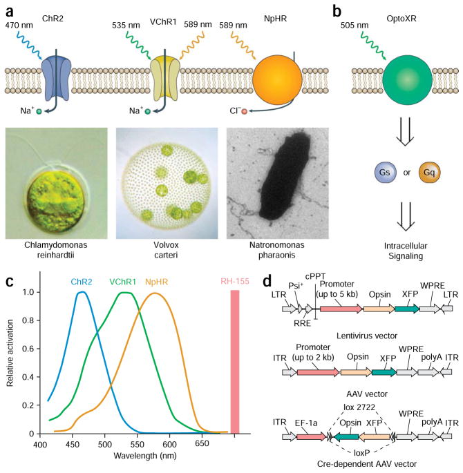
Elucidation of the neural substrates underlying complex animal behaviors depends on precise activity control tools, as well as compatible readout methods. Recent developments in optogenetics have addressed this need, opening up new possibilities for systems neuroscience. Interrogation of even deep neural circuits can be conducted by directly probing the necessity and sufficiency of defined circuit elements with millisecond-scale, cell type-specific optical perturbations, coupled with suitable readouts such as electrophysiology, optical circuit dynamics measures and freely moving behavior in mammals. Here we collect in detail our strategies for delivering microbial opsin genes to deep mammalian brain structures in vivo, along with protocols for integrating the resulting optical control with compatible readouts (electrophysiological, optical and behavioral). The procedures described here, from initial virus preparation to systems-level functional readout, can be completed within 4-5 weeks. Together, these methods may help in providing circuit-level insight into the dynamics underlying complex mammalian behaviors in health and disease.


Figures

Figure 1
Figure 1
Optogenetic tools. (a) Naturally occurring light-responsive effectors and their microbial sources: ChR2 from Chlamydomonas reinhardtii, VChR1 from Volvox carteri and NpHR from Natronomonas pharaonis; useful light wavelengths for each are indicated. ChR2 and VChR1 are cation-conducting channels and NpHR is a chloride pump. (b) Engineered synthetic rhodopsins for optical control of well-defined intracellular biochemical signaling. The intracellular loops of bovine rhodopsin have been replaced with the intracellular loops of G protein-coupled receptors (GPCRs) to yield light-activated chimeric GPCRs. Green light illumination leads to activation of the downstream Gq and Gs signaling pathways. (c) Action spectra. The absorbance wavelength of the voltage-sensitive dye (VSD) RH 155 is sufficiently separated from the light-sensitive range of all rhodopsins, therefore making it possible to integrate VSD imaging with optogenetic modulation. (d) Viral vectors for introducing microbial opsin genes into the brain. Top and middle: Lentiviral and AAV vectors can be used to deliver a cell-specific promoter along with the opsin gene and its fluorescent marker. Bottom: Cre-dependent adeno-associated virus (AAV) vector carries a doubly floxed inverted opsin (DIO) fusion gene. Upon transduction into Cre recombinase-expressing cells, the opsin fusion gene will be irreversibly inverted and enable cell-specific gene expression. Part a was modified with permission from Nature (Nature © 2007; Macmillan Publishers Ltd.).
Figure 2
Figure 2
Stereotactic implantation of the cannula guide. (a) After mounting the animal into the stereotactic frame, a first incision is made to open the skin above the skull. The skin is gently pulled to the side to reveal the cranial sutures. (b) After quickly wiping the skull with hydrogen peroxide, the bregma and the lambda can be easily identified (marked spots). (c) A thin needle is used to align the skull. (d) A dental drill is used to create a small craniotomy at the desired location on the skull, without puncturing the dura. The dura is later removed using fine forceps to minimize damage to the cortex. (e) A cannula guide is implanted on the skull through the craniotomy. (f) Metabond and dental cement are used to secure the cannula guide to the skull. (g) Vetbond and sutures are used to close the incision around the cannulation site. (h) An internal cannula guide connected to a pump is inserted through the cannula guide and is used to infuse virus into the target area in the brain. (i) The animal is allowed to rest in a recovery cage after surgical implantation. The surgery was conducted according to established animal care guidelines and protocols at Stanford University.
Figure 3
Figure 3
Preparation of the optical fiber for in vivo neural control in mammals. (a) Diagram of the optical neural interface (ONI), consisting of a stereotactically implanted cannula, an optical fiber, and a solid state laser controlled by a signal generator. The fiber is prepared with the appropriate length to illuminate the target brain region. (b) The tools and parts (fiber with FC connection, dummy cannula, internal cannula, cannula guide, dental drill, super glue, diamond scribe and fiber stripping tool) needed to manufacture ONI fibers. (c,d) A drill is used to produce a small bore on the center of a cannula cap. (e,f) The steel tubing is removed from an internal cannula. The plastic adapter is used as the fiber guide. (g) A fiber stripping tool is used to remove the plastic cladding from a fiber to reveal the fiber core. (h) The bare fiber core is threaded through the bore on the cannula cap. (i,j) The plastic adapter from the internal cannula is also placed on the fiber. (k,l) Superglue is applied to the fiber to secure the plastic adapter against the plastic cladding on the fiber. The plastic adapter is held tightly against the fiber cladding for several minutes to allow the superglue to harden. (m) After superglue has hardened, the fiber is inserted through a cannula guide with the right projection length for the target brain region. (n) The cannula guide is securely clipped into the plastic adapter. (o) A diamond scribe is used to remove the excess fiber from the tip of the cannula guide. (p) The finished ONI fiber is allowed to protrude from the tip of the cannula guide by ~0.5 mm. Part a was modified with permission (Nature © 2007; Macmillan Publishers Ltd.).
Figure 4
Figure 4
Functional expression of microbial opsin genes in the rodent brain. (a) Fluorescence image superimposed on bright field image of the motor cortex of a wild-type mouse injected with 2 μl lentivirus carrying the CaMKIIα::ChR2–EYFP construct, into layer V of the anterior M1 cortical area (AP 2 mm; ML 2 mm; DV 2 mm); 2 weeks were allowed for expression before preparing 200 μm slices that were examined under confocal (images are z-projection of single planes). (b) The injection produced strong expression in layer V of the cortex (the dendrites projecting from layer V neurons to the surface of the brain are clearly visible). (c) Fibers from layer V of the cortex travel through the corpus callosum (CC) and striatum. (d) Axons terminating in target structures. (e) Optrode recording during optical stimulation of the subthalamic nucleus of a Thy-1::ChR2–EYFP mouse. Example trace shows ten pulses of 5 ms blue light (470 nm) flashes delivered at 20 Hz. At right, zoomed view shows a single 5 ms light flash and resulting evoked spike. (f) Fast all-optical interrogation: typical voltage sensitive dye imaging signal from an acute horizontal hippocampal slice stained with RH 155. Trace shows the voltage changes in the slice resulting from 10 pulses of 10 ms blue light flashes delivered at 20 Hz. Acquisition rate: 200 Hz. The trace is averaged over four acquisition periods and a region of interest of 147 μm × 133 μm was defined in the entorhinal cortex. (Scale bars: a, 1 mm; b, 500 μm; c, 50 μm; d, 25 μm.)

Similar articles

Cited by

References

    1. Boyden ES, Zhang F, Bamberg E, Nagel G, Deisseroth K. Millisecond-timescale, genetically targeted optical control of neural activity. Nat Neurosci. 2005;8:1263–1268. - PubMed
    1. Zhang F, Wang LP, Boyden ES, Deisseroth K. Channelrhodopsin-2 and optical control of excitable cells. Nat Methods. 2006;3:785–792. - PubMed
    1. Zhang F, et al. Red-shifted optogenetic excitation: a tool for fast neural control derived from Volvox carteri. Nat Neurosci. 2008;11:631–633. - PMC - PubMed
    1. Zhang F, et al. Multimodal fast optical interrogation of neural circuitry. Nature. 2007;446:633–639. - PubMed
    1. Airan RD, Thompson KR, Fenno LE, Bernstein H, Deisseroth K. Temporally precise in vivo control of intracellular signalling. Nature. 2009;458:1025–1029. - PubMed

Publication types