

"It's Not What You Say, But How You Say it": A Reciprocal Temporo-frontal Network for Affective Prosody
- PMID: 20204074
- PMCID: PMC2831710
- DOI: 10.3389/fnhum.2010.00019
"It's Not What You Say, But How You Say it": A Reciprocal Temporo-frontal Network for Affective Prosody
Abstract
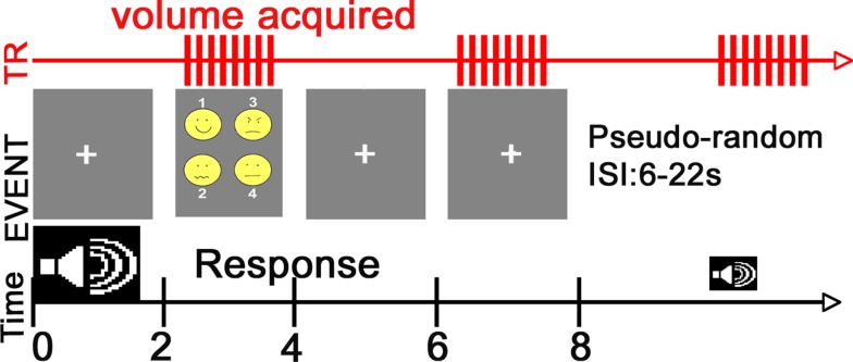
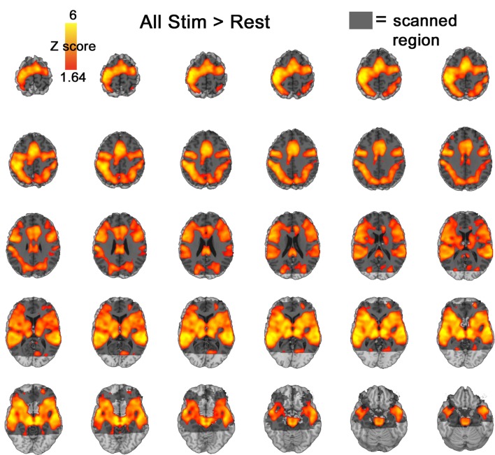
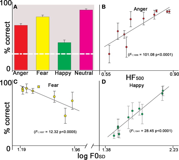
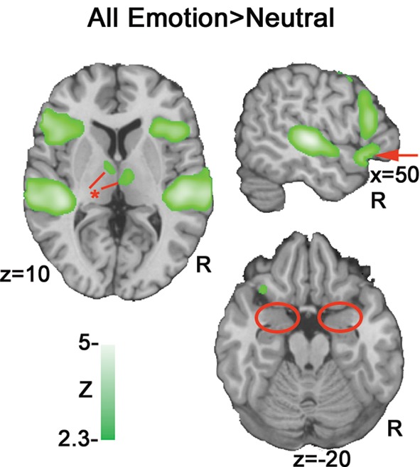
Humans communicate emotion vocally by modulating acoustic cues such as pitch, intensity and voice quality. Research has documented how the relative presence or absence of such cues alters the likelihood of perceiving an emotion, but the neural underpinnings of acoustic cue-dependent emotion perception remain obscure. Using functional magnetic resonance imaging in 20 subjects we examined a reciprocal circuit consisting of superior temporal cortex, amygdala and inferior frontal gyrus that may underlie affective prosodic comprehension. Results showed that increased saliency of emotion-specific acoustic cues was associated with increased activation in superior temporal cortex [planum temporale (PT), posterior superior temporal gyrus (pSTG), and posterior superior middle gyrus (pMTG)] and amygdala, whereas decreased saliency of acoustic cues was associated with increased inferior frontal activity and temporo-frontal connectivity. These results suggest that sensory-integrative processing is facilitated when the acoustic signal is rich in affective information, yielding increased activation in temporal cortex and amygdala. Conversely, when the acoustic signal is ambiguous, greater evaluative processes are recruited, increasing activation in inferior frontal gyrus (IFG) and IFG STG connectivity. Auditory regions may thus integrate acoustic information with amygdala input to form emotion-specific representations, which are evaluated within inferior frontal regions.
Keywords: amygdala; auditory cortex; emotion; inferior frontal gyrus; prosody; speech.
Figures


Similar articles
-
Specific brain networks during explicit and implicit decoding of emotional prosody.Cereb Cortex. 2012 May;22(5):1107-17. doi: 10.1093/cercor/bhr184. Epub 2011 Jul 12. Cereb Cortex. 2012. PMID: 21750247
-
Towards a fronto-temporal neural network for the decoding of angry vocal expressions.Neuroimage. 2012 Sep;62(3):1658-66. doi: 10.1016/j.neuroimage.2012.06.015. Epub 2012 Jun 19. Neuroimage. 2012. PMID: 22721630
-
Neural Tuning to Low-Level Features of Speech throughout the Perisylvian Cortex.J Neurosci. 2017 Aug 16;37(33):7906-7920. doi: 10.1523/JNEUROSCI.0238-17.2017. Epub 2017 Jul 17. J Neurosci. 2017. PMID: 28716965 Free PMC article.
-
Cerebral processing of linguistic and emotional prosody: fMRI studies.Prog Brain Res. 2006;156:249-68. doi: 10.1016/S0079-6123(06)56013-3. Prog Brain Res. 2006. PMID: 17015084 Review.
-
The brain basis of audiovisual affective processing: Evidence from a coordinate-based activation likelihood estimation meta-analysis.Cortex. 2019 Nov;120:66-77. doi: 10.1016/j.cortex.2019.05.016. Epub 2019 Jun 8. Cortex. 2019. PMID: 31255920 Review.
Cited by
-
Auditory emotion recognition impairments in schizophrenia: relationship to acoustic features and cognition.Am J Psychiatry. 2012 Apr;169(4):424-32. doi: 10.1176/appi.ajp.2011.11081230. Am J Psychiatry. 2012. PMID: 22362394 Free PMC article.
-
Neurofunctional correlates of expressed vocal affect in social phobia.Cogn Affect Behav Neurosci. 2011 Sep;11(3):413-25. doi: 10.3758/s13415-011-0032-3. Cogn Affect Behav Neurosci. 2011. PMID: 21509493
-
Auditory tasks for assessment of sensory function and affective prosody in schizophrenia.Compr Psychiatry. 2014 Nov;55(8):1862-74. doi: 10.1016/j.comppsych.2014.08.046. Epub 2014 Aug 19. Compr Psychiatry. 2014. PMID: 25214372 Free PMC article.
-
Structural and functional connectivity of the subthalamic nucleus during vocal emotion decoding.Soc Cogn Affect Neurosci. 2016 Feb;11(2):349-56. doi: 10.1093/scan/nsv118. Epub 2015 Sep 23. Soc Cogn Affect Neurosci. 2016. PMID: 26400857 Free PMC article.
-
Social cognitive deficits and their neural correlates in progressive supranuclear palsy.Brain. 2012 Jul;135(Pt 7):2089-102. doi: 10.1093/brain/aws128. Epub 2012 May 26. Brain. 2012. PMID: 22637582 Free PMC article.