. 2010:2010:289360.
doi: 10.1155/2010/289360. Epub 2010 Mar 3.

Neutrophils compromise retinal pigment epithelial barrier integrity

Affiliations

Neutrophils compromise retinal pigment epithelial barrier integrity

Jiehao Zhou et al. J Biomed Biotechnol. 2010.

Abstract

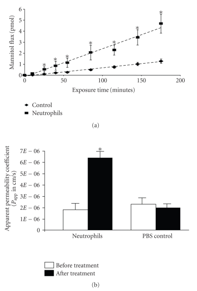
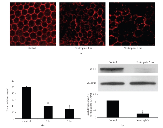
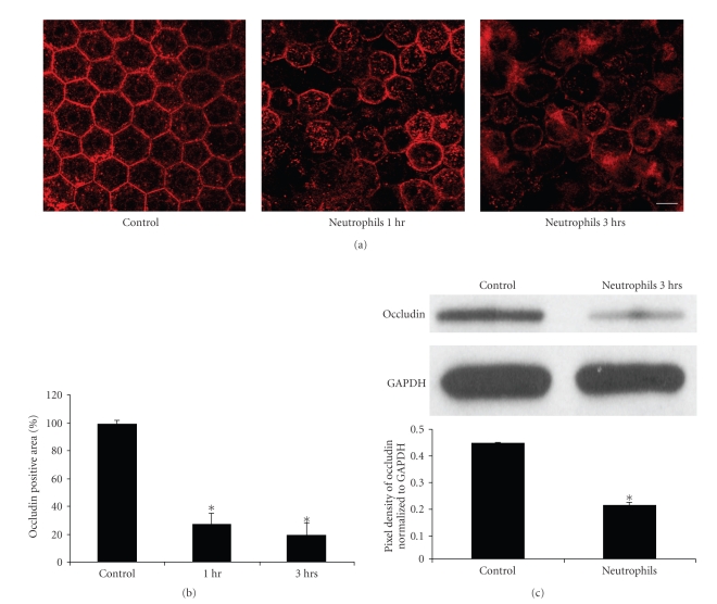
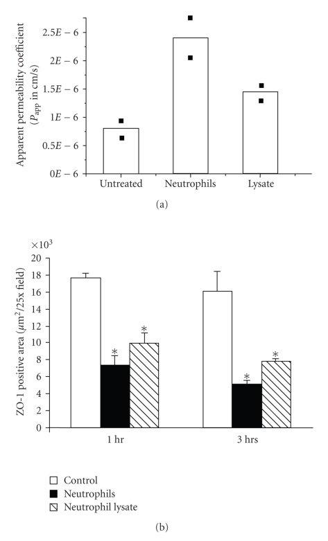
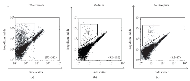
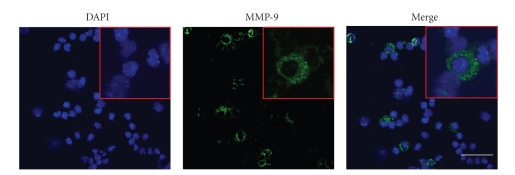
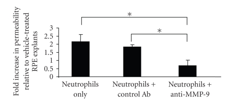
We hypothesized that neutrophils and their secreted factors mediate breakdown of the integrity of the outer blood-retina-barrier by degrading the apical tight junctions of the retinal pigment epithelium (RPE). The effect of activated neutrophils or neutrophil cell lysate on apparent permeability of bovine RPE-Choroid explants was evaluated by measuring [3H] mannitol flux in a modified Ussing chamber. The expression of matrix metalloproteinase- (MMP-) 9 in murine peritoneal neutrophils, and the effects of neutrophils on RPE tight-junction protein expression were assessed by confocal microscopy and western blot. Our results revealed that basolateral incubation of explants with neutrophils decreased occludin and ZO-1 expression at 1 and 3 hours and increased the permeability of bovine RPE-Choroid explants by >3-fold (P < .05). Similarly, basolateral incubation of explants with neutrophil lysate decreased ZO-1 expression at 1 and 3 hours (P < .05) and increased permeability of explants by 75%. Further, we found that neutrophils prominently express MMP-9 and that incubation of explants with neutrophils in the presence of anti-MMP-9 antibody inhibited the increase in permeability. These data suggest that neutrophil-derived MMP-9 may play an important role in disrupting the integrity of the outer blood-retina barrier.


Figures

Figure 1
Figure 1
Neutrophil exposure increased explant permeability. (a) The kinetics of RPE explant permeability following neutrophil treatment. The basolateral side of the explant was incubated with neutrophils for 1 to 3 hours at 37°C. (b) Neutrophil-induced increase of permeability of the RPE explant. The basolateral side of the explant was incubated with neutrophils for 3 hours at 37°C. The apparent permeability was determined by [3H]-mannitol flux. The increased permeability induced by neutrophils is significant compared with control. *represents P < .05. Addition of PBS to the explant did not change the permeability.
Figure 2
Figure 2
Neutrophils induced disruption of the tight junction protein ZO-1 in the RPE monolayer. (a) ZO-1 immunofluorescence staining on RPE-Choroidal explants. (b) Quantative analysis of ZO-1 immunofluorecence area on RPE–Choroidal explants. Bovine RPE-Choroidal explants were incubated with neutrophils for 1 to 3 hours at 37°C. ZO-1 expression was determined by confocal immunomicroscopy using anti-ZO-1 Ab. Quantitation of ZO-1 staining positive area was analyzed by using LSM 510 image software. *represents P < .01. Bar = 50 μm. (c) Western blot analysis of ZO-1 expression after RPE choroidal explants exposed to neutrophils for 3 hours. In Figure 2(c), the result of densitometry from the western blot (upper panel), normalized to GAPDH, shows significant loss of ZO-1 protein, P < .05.
Figure 3
Figure 3
Neutrophils induced disruption of tight junction protein occludin in the RPE monolayer. (a) Occludin immunofluorescence staining on RPE-Choroidal explants. (b) Quantative analysis of occludin immunofluorecence area on RPE–Choroidal explants. Bovine RPE-Choroidal explants were incubated with neutrophils for 1–3 hours at 37°C. Occludin expression was determined by confocal immunomicroscopy using anti-ZO-1 Ab. Quantitation of occludin staining positive area was analyzed by using LSM 510 image software. *represents P < .01.  Bar = 50 μm. (c) Western blot shows the effects of Neutrophils on occludin expression after 3-hour incubation. In (c), the result of densitometry from the western blot (upper panel), normalized to GAPDH, shows significant loss of occludin protein, P < .05.
Figure 4
Figure 4
Neutrophil and neutrophil lysate exposure increased the explant permeability (a) and decreased ZO-1 (b) expression. In (a), the basolateral side of the explant was incubated with neutrophils or neutrophil lysate for 3 hours at 37°C. The apparent permeability was determined by [3H]-mannitol flux. Increased permeability induced by neutrophils or neutrophil lysate is seen compared with control (two separate experiments); the variations between experiments were less than 10%. In (b), the disruption of tight junction protein ZO-1 by neutrophils or neutrophil lysate in the RPE monolayer was compared by confocal immunofluorescent staining in 3 separate experiments. Quantitation of ZO-1 staining positive area was determined by LSM 510 image software. Both neutrophil and neutrophil lysate decreased the expression of ZO-1 expression. *P < .05
Figure 5
Figure 5
The effects of neutrophil incubation on RPEs cell death. Human RPEs were incubated with neutrophils, C2-ceramide (20 μM; positive control), or medium (negative control) for 3 hours and then stained by Propidium iodide (PI). Cell death was evaluated by flow cytometry using 5000 cells for each experiment. The number of nonviable cells (R2) is indicated for each condition. C2-ceramide induced cell death (7.6%). Cells incubated with neutrophils or medium alone had lower cell death, namely, 1.7% and 2.0% cell death, respectively.
Figure 6
Figure 6
Localization of MMP-9 in mouse neutrophils determined by immunofluorescent confocal microscopy. DAPI, nuclear counterstained (blue); MMP-9 immunofluorescence (green). Representative neutrophils are shown at higher magnification in inset. Bar = 30 μm.
Figure 7
Figure 7
Anti-MMP-9 antibody attenuated neutrophil-mediated increase in permeability. The neutrophils were pre-treated with either anti-MMP-9 antibody or control antibody followed by incubation with explants for 3 hours at 37°C. The apparent permeability was determined by [3H]-mannitol flux and the data were presented as the fold increase in permeability relative to vehicle-treated samples. *represents P < .05.

References

    1. Campochiaro PA, Bryan JA, III, Conway BP, Jaccoma EH. Intravitreal chemotactic and mitogenic activity. Implication of blood-retinal barrier breakdown. Archives of Ophthalmology. 1986;104(11):1685–1687. - PubMed
    1. Jaccoma EH, Conway BP, Campochiaro PA. Cryotherapy causes extensive breakdown of the blood-retinal barrier. A comparison with argon laser photocoagulation. Archives of Ophthalmology. 1985;103(11):1728–2730. - PubMed
    1. Canataroglu H, Varinli I, Ozcan AA, Canataroglu A, Doran F, Varinli S. Interleukin (IL)-6, interleukin (IL)-8 levels and cellular composition of the vitreous humor in proliferative diabetic retinopathy, proliferative vitreoretinopathy, and traumatic proliferative vitreoretinopathy. Ocular Immunology and Inflammation. 2005;13(5):375–381. - PubMed
    1. Zheng M, Atherton SS. Cytokine profiles and inflammatory cells during HSV-1-induced acute retinal necrosis. Investigative Ophthalmology and Visual Science. 2005;46(4):1356–1363. - PubMed
    1. Thumann G, Hoffmann S, Hinton DR. Cell biology of the retinal pigment epithelium. In: Ryan S, editor. Retina. 4th edition. Vol. 1. London, UK: Elsevier; Mosby; 2005. pp. 137–152.

Publication types

LinkOut - more resources