. 2010 Feb 8:4:1.
doi: 10.3389/neuro.06.001.2010. eCollection 2010.

Three-dimensional reconstruction and segmentation of intact Drosophila by ultramicroscopy

Affiliations

Three-dimensional reconstruction and segmentation of intact Drosophila by ultramicroscopy

Nina Jährling et al. Front Syst Neurosci. .

Abstract

Genetic mutants are invaluable for understanding the development, physiology and behaviour of Drosophila. Modern molecular genetic techniques enable the rapid generation of large numbers of different mutants. To phenotype these mutants sophisticated microscopy techniques are required, ideally allowing the 3D-reconstruction of the anatomy of an adult fly from a single scan. Ultramicroscopy enables up to cm fields of view, whilst providing micron resolution. In this paper, we present ultramicroscopy reconstructions of the flight musculature, the nervous system, and the digestive tract of entire, chemically cleared, drosophila in autofluorescent light. The 3D-reconstructions thus obtained verify that the anatomy of a whole fly, including the filigree spatial organization of the direct flight muscles, can be analysed from a single ultramicroscopy reconstruction. The recording procedure, including 3D-reconstruction using standard software, takes no longer than 30 min. Additionally, image segmentation, which would allow for further quantitative analysis, was performed.

Keywords: Drosophila; flight muscle; imaging; light sheet microscopy; morphology; phenotyping; segmentation; ultramicroscopy.


Figures

Figure 1
Figure 1
Standard configuration of an ultramicroscopy setup. A laser beam is expanded and homogenized by two convex lenses and then split into two separate light pathways. Both beams are focussed by cylindrical lenses and slit apertures (1), forming a thin light sheet. The light sheet illuminates the specimen placed in a transparent specimen chamber (2) filled with clearing solution. Hence, fluorescence is only exhibited in those parts of the specimen, which are in the focal plane. The fluorescence image is projected to a camera target using a microscope objective, and the excitation light is blocked by a matched optical band pass filter. By stepping the specimen chamber vertically through the light sheet using a computer controlled jack (3), a stack of optical slices is generated.
Figure 2
Figure 2
White eyed Drosophila, placed on a Siemens star after dehydration and clearing. The fly is nearly completely translucent. Length of scale bar 1 mm.
Figure 3
Figure 3
(A) Reconstructed surface of an entire fly. Scale bar 100 μm. (B) Sagittal view of the fly's inner anatomy, showing parts of the flight muscles, the nervous, and the cardiac system. DLMs, dorso longitudinal muscles; ThAGl, thoracico-abdominal ganglion; PV, proventriculus; CB, cibarium. Scale bar: 100 μm. (C) Detail of the fly virtually sectioned along a transversal plane through the thorax. DVM-I, dorsal–ventral muscles; SGs, salivary glands. Scale bar: 100 μm. (D) Detail showing the direct flight muscles DFM49–DFM56. DFM52 is only rudimentarily visible, because it is clipped by the viewing plane. Scale bar 40 μm.
Figure 4
Figure 4
Cross sections in three orthogonal directions (A–C) were used to segment anatomical structures in three different orientations. ES, oesophagus; CN, cervical connective; DLMs, dorsal longitudinal muscles; DVM-I/-II/-III, dorsal–ventral muscles; PV, proventriculus; TDT, tergal depressor of the trochanter; ThAGl, thoracico-abdominal ganglion. I: Sagittal plane, II: transversal plane, III: coronal plane. (A) Sagittal optical slice. (B) Computed coronal slice. (C) Computed transversal slice. Length of scale bars 100 μm.
Figure 5
Figure 5
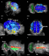
Segmentation of Drosophila organs. (A,B) Overview of the entire fly, illustrating major components of the nervous system, the intestinal tract, and the musculature. (C,D) Detail of the flight musculature. DLM, dorsal longitudinal muscles (dark-blue); DVM-I/II/III, dorsal–ventral muscles (blue); DFM49–DFM54, direct flight muscles (yellow); TDT, tergal depressor of the trochanter (light-blue). (E-F) Detail of the CNS and the intestinal system. Brain (dark green), CN, cervical connective (green); ThAGl, thoracico-abdominal ganglion, (light-green); CB, cibarium (light-rose); ES, oesophagus (rose); PV, proventriculus (red); SGs, pair of salivary glands (brown); CR, crop (orange). Length of scale bar 200 μm.

References

    1. Becker K., Jährling N., Kramer E. R., Schnorrer F., Dodt H.-U. (2008). Ultramicroscopy: 3D-reconstruction of large microscopic specimens. J. Biophoton. 1, 36–4210.1002/jbio.200710011 - DOI - PubMed
    1. Dietzl G., Chen D., Schnorrer F., Su K.-C., Barinova Y., Fellner M., Gasser B., Kinsey K., Oppel S., Scheiblauer S., Couto A., Marra V., Keleman K., Dickson B. J. (2007). A genome-wide transgenic RNAi library for conditional gene inactivation in Drosophila. Nature 448, 151–15610.1038/nature05954 - DOI - PubMed
    1. Dodt H.-U., Leischner U., Schierloh A., Jährling N., Mauch C.P., Deininger K., Deusing J.M., Eder M., Zieglgänsberger W., Becker K. (2007). Ultramicroscopy: three-dimensional visualization of neural networks in the whole mouse brain. Nat. Methods 4, 331–33610.1038/nmeth1036 - DOI - PubMed
    1. Dutta D., Anant S., Ruiz-Gomez M., Bate M., VijayRaghavan K. (2004). Founder myoblasts and fibre number during myogenesis in Drosophila. Development 131, 3761–377210.1242/dev.01249 - DOI - PubMed
    1. Ghazi A., Anant S., VijayRaghavan K. (2000). Apterous mediates development of direct muscles autonomously and indirect flight muscles through epidermal cues. Development 127, 5309–5318 - PubMed

LinkOut - more resources