. 2010 Jun;67(12):2025-38.
doi: 10.1007/s00018-010-0308-8. Epub 2010 Mar 4.

Tubulin chaperone E binds microtubules and proteasomes and protects against misfolded protein stress

Affiliations

Tubulin chaperone E binds microtubules and proteasomes and protects against misfolded protein stress

Olga Voloshin et al. Cell Mol Life Sci. 2010 Jun.

Abstract

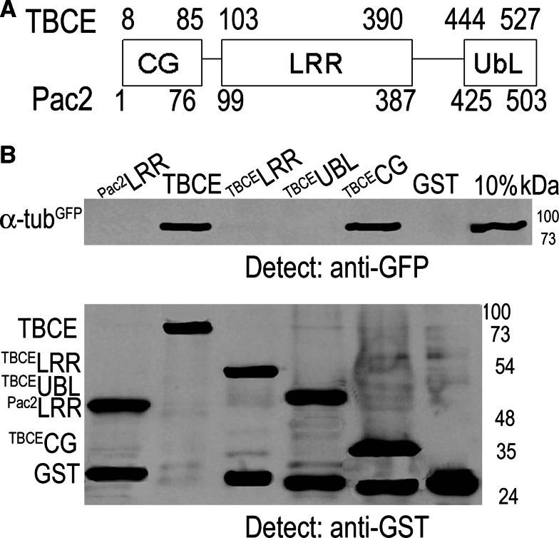
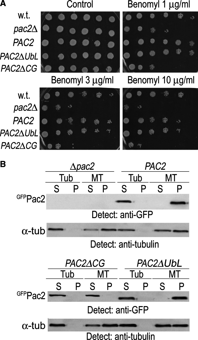
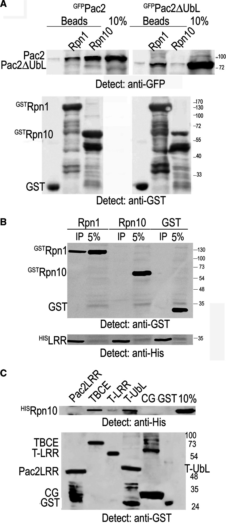
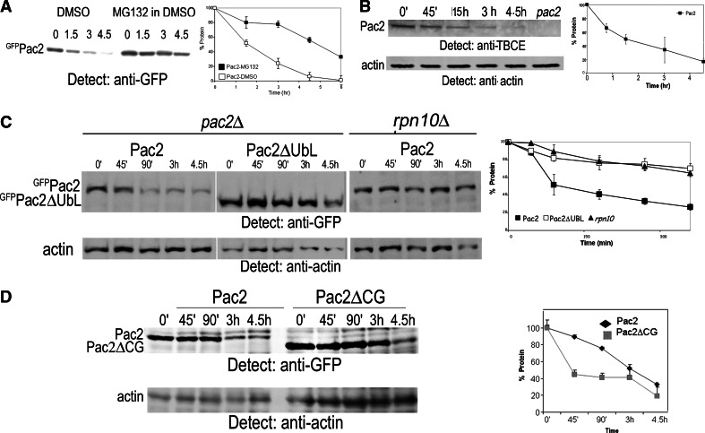
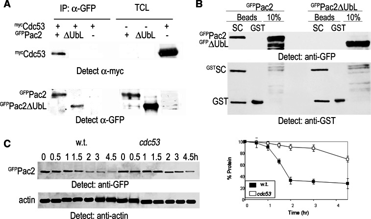
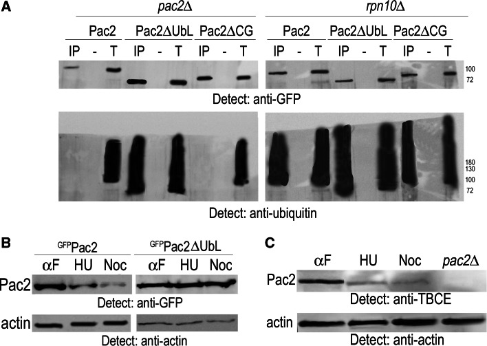
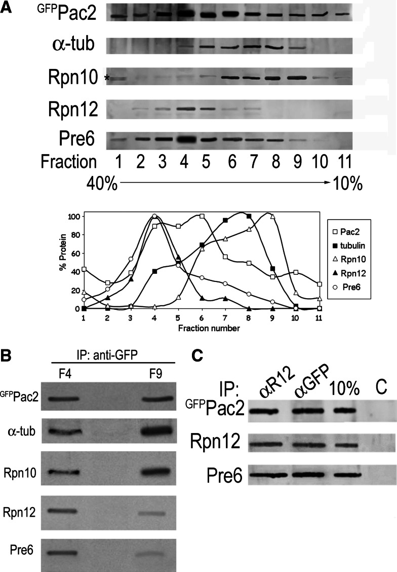
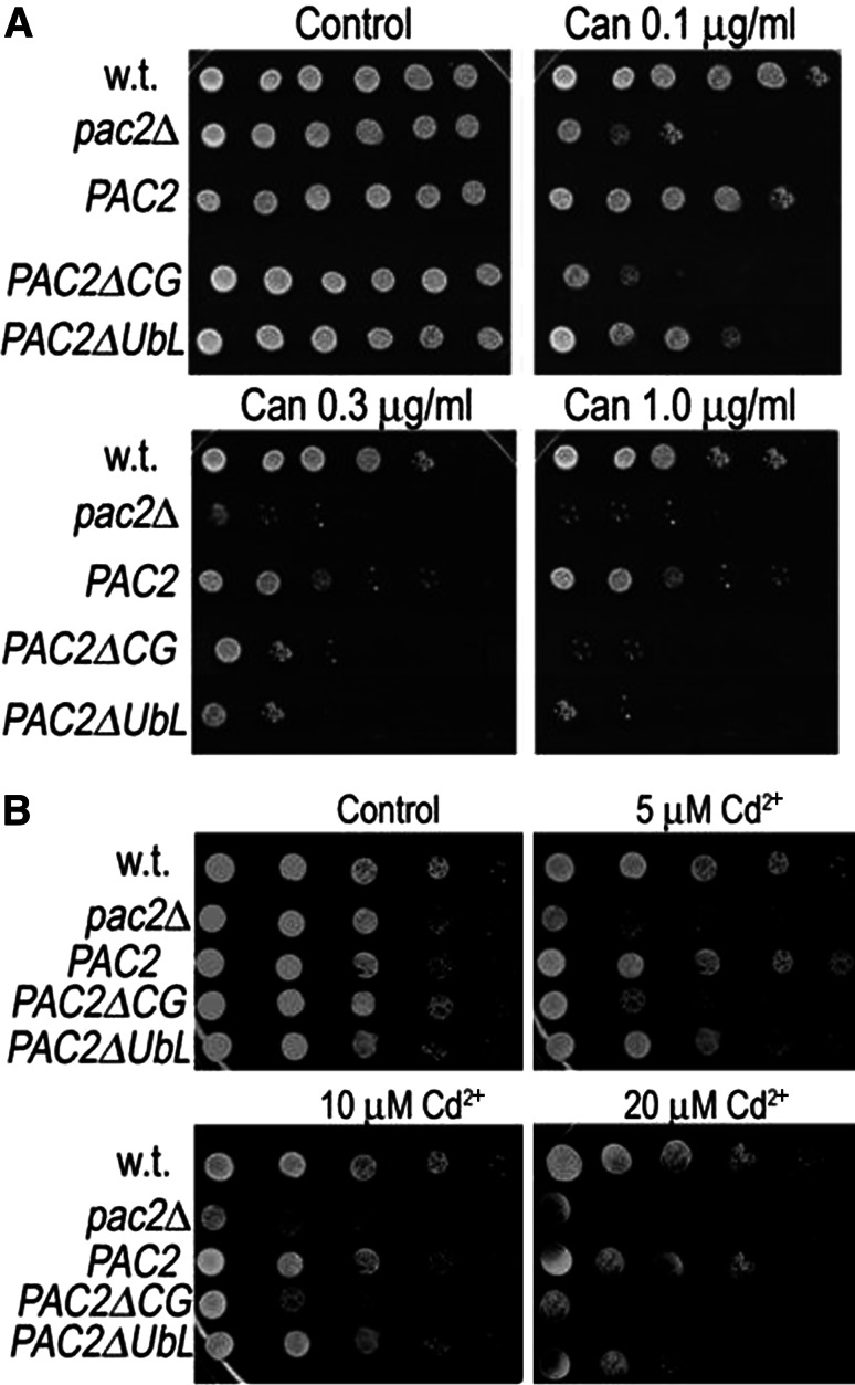
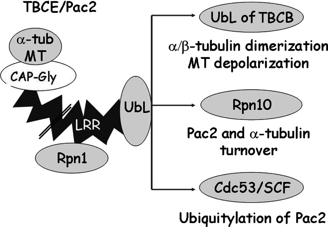
Mutation of tubulin chaperone E (TBCE) underlies hypoparathyroidism, retardation, and dysmorphism (HRD) syndrome with defective microtubule (MT) cytoskeleton. TBCE/yeast Pac2 comprises CAP-Gly, LRR (leucine-rich region), and UbL (ubiquitin-like) domains. TBCE folds alpha-tubulin and promotes alpha/beta dimerization. We show that Pac2 functions in MT dynamics: the CAP-Gly domain binds alpha-tubulin and MTs, and functions in suppression of benomyl sensitivity of pac2Delta mutants. Pac2 binds proteasomes: the LRR binds Rpn1, and the UbL binds Rpn10; the latter interaction mediates Pac2 turnover. The UbL also binds the Skp1-Cdc53-F-box (SCF) ubiquitin ligase complex; these competing interactions for the UbL may impact on MT dynamics. pac2Delta mutants are sensitive to misfolded protein stress. This is suppressed by ectopic PAC2 with both the CAP-Gly and UbL domains being essential. We propose a novel role for Pac2 in the misfolded protein stress response based on its ability to interact with both the MT cytoskeleton and the proteasomes.


Figures

Fig. 1
Fig. 1
The CAP-Gly domain mediates interaction of TBCE with α-tubulin. a Domain structure of human TBCE and yeast Pac2 showing CAP-Gly (CG), LRR, and UbL domains based on [11]. b Yeast extract from cells with tagged genomic α-tubulinGFP was incubated with GSH beads to which were bound recombinant yeast GSTPac2-LRR domain, human GSTTBCE, GSTTBCE-LRR, GSTTBCE-UbL, GSTTBCE-CG, or GST, produced in bacteria. The bead fraction was analyzed by Western blotting with anti-GFP and anti-GST antibodies; 10% is an aliquot of the yeast extract incubated with the beads
Fig. 2
Fig. 2
Pac2 affects MT dynamics. a Transformed pac2Δ mutants expressing pGAL-GFP-PAC2, pGAL-GFP-PAC2ΔUBL, or pGAL-GFP-PAC2ΔCAP-GLY (CG) were induced overnight with galactose and the next morning were observed in the fluorescent microscope to ascertain production of each protein. Serial threefold dilutions of wild-type, pac2Δ mutants, or pac2Δ mutants expressing pGAL-GFP-PAC2, pGAL-GFP-PAC2ΔUBL, or pGAL-GFP-PAC2ΔCG were spotted onto plates without (control) or with 1, 3, or 10 μg/ml benomyl and grown for 48 h prior to documentation. b Tubulin heterodimers (Tub) or microtubules polymerized in vitro (MT) were mixed with yeast extracts from pac2Δ cells or from pac2Δ cells producing GFPPac2, GFPPac2ΔUbL, or GFPPac2ΔCG and spun through a glycerol cushion (“Materials and methods”). The supernatant (S) and pellet (P) fractions were analyzed separately by Western blotting for the presence of GFPPac2, GFPPac2ΔUbL, and GFPPac2ΔCG
Fig. 3
Fig. 3
The LRR domain interacts with Rpn1 of the 19S RP. a GSH beads to which recombinant yeast GSTRpn1, GSTRpn10, or GST was bound were incubated with yeast extracts from pac2Δ mutants expressing pGAL-GFP-PAC2 or pGAL-GFP-PAC2ΔUBL. The bead fraction was analyzed by Western blotting with anti-GFP and anti-GST antibodies; 10% is the volume of total cell extract used in the experiment. b Recombinant yeast Pac2 HisLRR domain produced in bacteria was bound to nickel beads and incubated with a bacterial lysate from cells that produced yeast GSTRpn1, GSTRpn10, or GST. The bead fraction was analyzed by Western blotting first with anti-GST and then with anti-His antibodies; 5% is of the bacterial cell lysate. c Recombinant yeast HISRpn10 produced in bacteria was incubated with yeast GSTPac2-LRR domain, full-length human TBCE, or subclones of the TBCE LRR (T-LRR), UbL (T-UbL), or CAP-Gly (CG) domains, or with control GST beads, all produced in bacteria. HISRpn10 that bound TBCE protein was detected with anti-His antibodies; the proteins on the beads are shown using anti-GST antibodies, and 10% is an aliquot of HISRpn10 bacterial lysate incubated with the beads
Fig. 4
Fig. 4
Turnover of Pac2 depends on interaction of its UbL domain with Rpn10. a Pac2 is stabilized in cells treated with the proteasome inhibitor MG132. pGAL-GFP-PAC2 transformed into pdr5Δ mutant cells was induced overnight with 2% galactose. Next morning the cells were diluted 1:3, and after 2 h 100 μl MG132 dissolved in DMSO was added (controls were treated with DMSO), together with 3% glucose to repress transcription and 10 mM CHX to inhibit translation. Equal aliquots of cells were analyzed at the zero time point and at the times indicated above each lane by anti-GFP immunoprecipitation followed by Western blotting. Graph shows protein remaining at each time point. b Half-life of endogenous Pac2 determined using an affinity-purified polyclonal antibody to human TBCE; 10 mM CHX was added at the zero time point, and equal aliquots were analyzed by TCA precipitation and Western blotting at the times indicated. Extract from pac2Δ mutants was used as a control for the specificity of the antibody; anti-actin was used as a loading control. c Pac2 and Pac2ΔUbL turnover in pac2Δ and rpn10Δ cells: pGAL-GFP-PAC2 or pGAL-GFP-PAC2ΔUBL were induced overnight in pac2Δ mutants, and pGAL-GFP-PAC2 was induced overnight in rpn10Δ mutants. At the zero time point, 3% glucose and 10 mM CHX were added, and equal aliquots of cells were TCA-precipitated for Western blotting at the times indicated above the lanes. Actin was used as a loading control. d Pac2 and Pac2ΔCG turnover in pac2Δ mutants: experimental conditions as in c
Fig. 5
Fig. 5
The Pac2 UbL domain interacts with the SCF complex. a GFPPac2 and GFPPac2ΔUbL were immunoprecipitated with anti-GFP from yeast extracts made from cells that expressed mycCdc53. The bead fractions were blotted with anti-myc antiserum to detect Cdc53 and with anti-GFP to detect immunoprecipitated GFPPac2. b GSTSkp1-Cdc53 heterodimers (SC) from Sf9 cells on GSH beads or GST peptides were incubated with yeast extracts from pac2Δ mutants expressing pGFP-PAC2 or pGFP-PAC2ΔUBL. Interaction was detected by Western blot with anti-GFP antibodies. c Half-life of Pac2 in wild-type cells and in cdc53 mutants measured at 37°C. Experimental details as in Fig. 4c
Fig. 6
Fig. 6
The Pac2 CAP-Gly and UbL domains are not required for ubiquitylation. a Pac2, Pac2ΔUbL, and Pac2ΔCAP-Gly (CG) were produced in pac2Δ and rpn10Δ mutants and immunoprecipitated with anti-GFP antibodies and Protein A beads (IP) or without antibodies (−). They were analyzed by Western blotting with anti-GFP and anti-ubiquitin antibodies. T is 5% of the extract used. b Ectopic Pac2 and Pac2ΔUbL levels at different stages of the cell cycle. pdr5Δ mutants expressing pGAL-GFP-PAC2 or pGAL-GFP-PAC2ΔUBL were arrested in G1 with α-factor, in S phase with HU, and in prometaphase with nocodazole. Equal aliquots were precipitated with TCA and blotted with anti-GFP and anti-actin antibodies. The ratio of Pac2 and Pac2ΔUbL to the actin loading control is shown in the histogram. c Levels of endogenous Pac2 in wild-type cells were detected with a rabbit polyclonal antibody to human TBCE in cells arrested in different stages of the cell cycle as above. pac2Δ mutants are shown as a control for the specificity of the human antibody
Fig. 7
Fig. 7
Pac2 complexes in vivo. a Western analysis of 11 40–10% glycerol gradient fractions blotted with anti-GFP antibodies to detect GFPPac2 (MW 89 KDa); and with anti-α-tubulin (MW 50 KDa); with anti-Rpn10 (MW 30 KDa) and anti-Rpn12 (MW 32 KDa) to detect the 19S RP; and with anti-Pre6 (MW 28.5 KDa) antibodies to detect the 20S CP. Graphs show relative amount of each protein in the different fractions. b Comparison of the Pac2 complexes from Fraction 4 and Fraction 9. GFPPac2 was immunoprecipitated with anti-GFP antibodies, and the bead fraction was separated by SDS-PAGE for Western blotting with anti-GFP, anti-α-tubulin, anti-Rpn10, anti-Rpn12, and anti-Pre6 antibodies. c Immunoprecipitation of aliquots of Fraction 4 with anti-Rpn12 (αR12 lane) to immunoprecipitate the 19S RP or anti-GFP antibodies (αGFP lane) to immunoprecipitate GFPPac2. Components of the complex that co-immunoprecipitated in each fraction were detected by Western blotting with the antibodies used in Fig. 8a. Controls (C) were Protein A beads without antibody; 10% is a TCA precipitation of an aliquot of the gradient fraction
Fig. 8
Fig. 8
pac2Δ mutants are sensitive to canavanine and to cadmium stresses. a Suppression of canavanine sensitivity of pac2Δ mutants by ectopic PAC2. Serial threefold dilutions of wild-type, pac2Δ mutants, or pac2Δ mutants expressing pGAL-GFP-PAC2, pGAL-GFP-PAC2ΔCAP-Gly (CG), or pGAL-GFP-PAC2ΔUbL were plated on cells with different concentrations of canavanine as indicated above the plates. They were photographed after 48 h at 30oC. b Suppression of cadmium sensitivity of pac2Δ mutants by ectopic PAC2. Cells were spotted as above onto plates with different concentrations of cadmium chloride as indicated above the plates
Fig. 9
Fig. 9
Interactions of the Pac2 protein domains. Left Pac2/TBCE protein domains. The CAP-Gly domain interacts with α-tubulin and MTs; the LRR domain interacts with Rpn1; the UbL domain: a dimerizes with the UbL of TBCB [16, 18] and affects MT dynamics; b interacts with Rpn10 and this determines turnover of Pac2 (present study), and of α-tubulin[52]; c interacts with the SCF ubiquitin ligase complex core subunits, Skp1-Cdc53 (present study). We propose that by engaging in these different interactions the UbL domain could fulfill a regulatory role in MT dynamics. The LRR domain is not drawn to scale (double line)

Similar articles

Cited by

References

    1. Parvari R, Hershkovitz E, Grossman N, Gorodischer R, Loeys B, Zecic A, Mortier G, Gregory S, Sharony R, Kambouris M, Sakati N, Meyer BF, Al Aqeel AI, Al Humaidan AK, Al Zanhrani F, Al Swaid A, Al Othman J, Diaz GA, Weiner R, Khan KT, Gordon R, Gelb BD. Mutation of TBCE causes hypoparathyroidism-retardation-dysmorphism and autosomal recessive Kenny-Caffey syndrome. Nat Genet. 2002;32:448–452. doi: 10.1038/ng1012. - DOI - PubMed
    1. Hoyt MA, Macke JP, Roberts BT, Geiser JR. Saccharomyces cerevisiae PAC2 functions with CIN1, 2 and 4 in a pathway leading to normal microtubule stability. Genetics. 1997;146:849–857. - PMC - PubMed
    1. Radcliffe PA, Hirata D, Vardy L, Toda T. Functional dissection and hierarchy of tubulin-folding cofactor homologues in fission yeast. Mol Biol Cell. 1999;10:2987–3001. - PMC - PubMed
    1. Tian G, Lewis SA, Feierbach B, Stearns T, Rommelaere H, Ampe C, Cowan NJ. Tubulin subunits exist in an activated conformational state generated and maintained by protein cofactors. J Cell Biol. 1997;138:821–832. doi: 10.1083/jcb.138.4.821. - DOI - PMC - PubMed
    1. Feierbach B, Nogales E, Downing KH, Stearns T. Alf1p, a CLIP-170 domain-containing protein, is functionally and physically associated with alpha-tubulin. J Cell Biol. 1999;144:113–124. doi: 10.1083/jcb.144.1.113. - DOI - PMC - PubMed

Publication types

MeSH terms

LinkOut - more resources