Review
. 2010 Jul;67(14):2343-62.
doi: 10.1007/s00018-010-0303-0. Epub 2010 Mar 5.

Biogenesis of bacterial inner-membrane proteins

Affiliations
Review

Biogenesis of bacterial inner-membrane proteins

Sandra J Facey et al. Cell Mol Life Sci. 2010 Jul.

Abstract

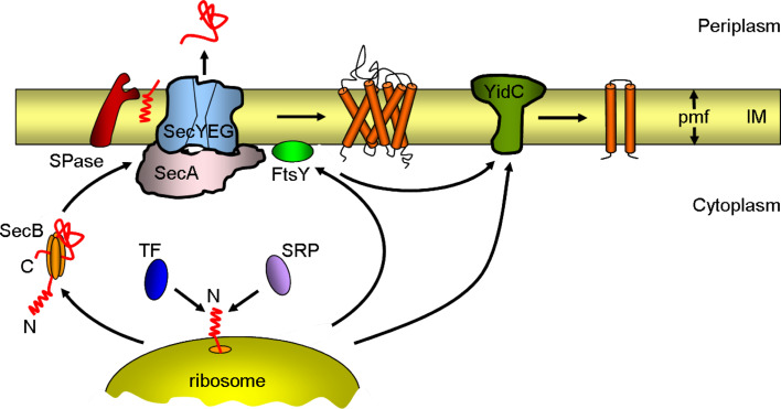
All cells must traffic proteins into and across their membranes. In bacteria, several pathways have evolved to enable protein transfer across the inner membrane, the periplasm, and the outer membrane. The major route of protein translocation in and across the cytoplasmic membrane is the general secretion pathway (Sec-pathway). The biogenesis of membrane proteins not only requires protein translocation but also coordinated targeting to the membrane beforehand and folding and assembly into their protein complexes afterwards to function properly in the cell. All these processes are responsible for the biogenesis of membrane proteins that mediate essential functions of the cell such as selective transport, energy conversion, cell division, extracellular signal sensing, and motility. This review will highlight the most recent developments on the structure and function of bacterial membrane proteins, focusing on the journey that integral membrane proteins take to find their final destination in the inner membrane.


Figures

Fig. 1
Fig. 1
Schematic overview of bacterial inner-membrane protein biogenesis. Newly synthesized proteins are targeted to the Sec complex either by the signal recognition particle (SRP) as soon as they emerge from the ribosome tunnel (co-translational translocation, mainly inner-membrane proteins) or by the tetrameric SecB chaperone after translation (post-translational translocation, mainly secretory and outer-membrane proteins). Trigger factor (TF) competes with SRP for the binding of the nascent protein. Proteins destined for the inner (IM) or outer membrane are transported into or across the inner membrane through the Sec complex. The complex consists of the SecYEG protein-conducting channel and the ATPase motor SecA. The signal peptidase (SPase) cleaves the signal sequence from preproteins at the outer face of the inner membrane. A few membrane proteins insert into the inner membrane via YidC. For simplicity, SecYEG is shown without its accessory components (SecDFYajC and YidC). pmf proton motive force
Fig. 2
Fig. 2
Schematic representation of the three-domain structure of Ffh and FtsY of E. coli. The domains are indicated with different colors: N domain purple, G domain yellow, M domain green, and A domain light blue. The five consensus elements responsible for GTP binding and hydrolysis (G1–G5) are highlighted in red, whereas the I-box and the inter-domain communication motifs (ALLEADV, DARGG, GQ) are indicated in blue. The two lipid-binding sites of FtsY are shown in grey
Fig. 3
Fig. 3
SecA-mediated protein translocation through the SecY pore. The scheme shows a model for the different steps in translocation. 1 SecA binds to a polypeptide substrate bearing an N-terminal signal sequence. 2 SecA opens toward SecY and transfers the signal sequence and early mature region of the polypeptide into SecY with the help of the two-helix finger. The polypeptide inserts as a loop structure. This step requires ATP hydrolysis. The insertion of the signal sequence of the polypeptide into SecY causes the plug to move away from the pore, which leads to a fully activated channel. 3 For further translocation, SecA releases the polypeptide into the pore and the helix finger is pulled back and reset. 4 SecA binds the next section of the polypeptide and moves toward the translocationally active SecY. 5 SecA then pushes the next section of the polypeptide into the pore by the helix finger. The signal sequence remains stationary, while the mature part of the polypeptide passes through the pore. These steps are coupled to ATP hydrolysis cycles and steps 35 are repeated until the polypeptide is fully translocated (not shown). For simplicity, SecA and SecYEG are shown as monomers and the initial stages of the process with SecB are not shown. The thick black line represents the polypeptide and the red rectangle represents the N-terminal signal sequence
Fig. 4
Fig. 4
Membrane topology and function of lactose permease (LacY) of E. coli. a Topological organization of LacY in E. coli membranes with (+PE) and without (−PE) phosphatidylethanolamine (PE). LacY consists of 12 transmembrane helices divided into two six-helix bundles. In the absence of PE (−PE), the N-terminal six-helix bundle with their associated extramembrane domains adopts an inverted topology with respect to the membrane bilayer, whereas the C-terminal six-helix bundle, except for TM VII, retains its native topology. The topological organization of TM VII in −PE cells remains unknown. The transmembrane helices are labeled in roman numerals consecutively from the N-(NH2) to C-(COOH) terminus. The cytoplasmic (green) and periplasmic (pink) extramembrane domains are indicated. b Schematic representation of conformational changes in LacY upon sugar binding. LacY uses a symport mechanism to couple the transport of lactose and H+ across the membrane. The proposed mechanism of transport alternates between the outward-facing conformation (exposed to the periplasmic side) shown in steps 1–3 and the inward-facing conformation (exposed to the cytoplasm) shown in steps 4–6. Starting from the outward-facing conformation 1 LacY is protonated 2 prior to substrate binding. 3 The binding of substrate to the binding site probably induces a conformation change in the two halves of the protein (N- and C-terminal six-helix bundles) that results in the inward-facing conformation. 4 The substrate is then released into the cytoplasm 5 followed by release of the H+. 6 After releasing the H+ inside, LacY returns to the outward-facing conformation 1. The N- and C-terminal six-helix bundles of LacY are colored light and dark blue, respectively. Substrate and H+ are represented by orange circles and green squares, respectively
Fig. 5
Fig. 5
Model of E. coli F1Fo-ATPase. The ATP synthase consists of two subcomplexes with eight different proteins, the membrane-integral Fo complex (a 1 b 2 c 10) and the peripheral F1 complex (α 3 β 3 γδε). The c subunit of Fo is linked to the γ and ε subunits to form the central rotor (purple). Subunits b and δ form a stator which ensures that subunits a in Fo and the α 3 β 3 hexamer of F1 do not rotate with the central rotor (γεc ring). The proton pathway lies between the a and c subunits. Either H+ translocation through Fo or ATP hydrolysis in F1 leads to the rotary movement of the central rotor element (γεc ring)
Fig. 6
Fig. 6
Schematic drawing of inward- and outward-facing structures of the maltose transporter, MalFGK2, of E. coli. The maltose transporter complex is composed of a periplasmic maltose-binding protein (MalE), two integral membrane proteins, MalF and MalG, and two copies of the cytoplasmic ATP-binding cassette subunit MalK. In the transport cycle, MalE binds and delivers maltose to the resting state transporter in the closed conformation (shown on the left). Interactions with MalE in the presence of maltose stimulate ATPase activity of the transporter by triggering major conformational changes in which the two cytosolic MalK subunits close and the transmembrane helices of MalF and MalG reorient to receive the substrate from MalE (shown in the middle). After ATP hydrolysis, the outward-facing conformation is no longer stable and the transporter returns to the inward-facing resting state releasing maltose (shown on the right). Maltose is indicated by a red sphere
Fig. 7
Fig. 7
A schematic representation of a bacterial flagellum basal body. The flagellum, which works as a rotary motor, consists of a motor component called the basal body, which spans from the cytoplasm to the outer membrane (OM), a flexible hook, and a filament. The basal body is composed of a rotor, a rod, a bushing, and a switch regulator. The core of the rotor is made up of FliF. The rod components are composed of four proteins, FlgB, FlgC, FlgF, and FlgG, and a rod adapter protein, FliE. The outer ring structures assembled around the rod consists of FlgI and FlgH and are thought to act as a molecular bushing of the rotary axial structure of the flagellum. The direction of rotation is controlled by the switch proteins, FliG, FliM and FliN. The stator part of the motor is composed of MotA and MotB, which span the inner membrane (IM). MotB is anchored to the peptidoglycan layer with its C-terminal periplasmic domain. Together both proteins form the proton pathway that powers the flagellum motor. The flagellar secretion apparatus (T3SS) consists of six integral membrane proteins FlhA, FlhB, FliO, FliP, FliQ, and FliR, and three soluble components, FliH, FliI, and FliJ, at the cytoplasmic face of the flagellar basal body. These proteins facilitate the export of flagellar substrates and are dependent on ATP hydrolysis by the ATPase FliI protein. The basal body of the flagellum is linked to a flexible hook consisting of FlgE and to a helical filament (FliC). The flagellum can rotate in either a clockwise or a counter-clockwise (arrow) direction

Similar articles

Cited by

References

    1. Kramer G, Rauch T, Rist W, Vorderwülbecke S, Patzelt H, Schulze-Specking A, Ban N, Deuerling E, Bukau B. L23 protein functions as a chaperone docking site on the ribosome. Nature. 2002;419:171–174. - PubMed
    1. Kaiser CM, Chang HC, Agashe VR, Lakshmipathy SK, Etchells SA, Hayer-Hartl M, Hartl FU, Barral JM. Real-time observation of trigger factor function on translating ribosomes. Nature. 2006;444:455–460. - PubMed
    1. Rutkowska A, Mayer MP, Hoffmann A, Merz F, Zachmann-Brand B, Schaffitzel C, Ban N, Deuerling E, Bukau B. Dynamics of trigger factor interaction with translating ribosomes. J Biol Chem. 2008;283:4124–4132. - PubMed
    1. Ferbitz L, Maier T, Patzelt H, Bukau B, Deuerling E, Ban N. Trigger factor in complex with the ribosome forms a molecular cradle for nascent proteins. Nature. 2004;431:590–596. - PubMed
    1. Bukau B, Deuerling E, Pfund C, Craig EA. Getting newly synthesized proteins into shape. Cell. 2000;101:119–122. - PubMed

Publication types

MeSH terms

Substances

LinkOut - more resources