Comparative Study
. 2010 Sep;468(9):2410-8.
doi: 10.1007/s11999-010-1283-y. Epub 2010 Mar 4.

Morphologic evaluation of chronic radial head dislocation: three-dimensional and quantitative analyses

Affiliations
Comparative Study

Morphologic evaluation of chronic radial head dislocation: three-dimensional and quantitative analyses

Kunihiro Oka et al. Clin Orthop Relat Res. 2010 Sep.

Abstract

Background: Treatment of chronic radial head dislocation is controversial, considering whether to reduce and reconstruct the proximal radioulnar joint. The anatomic alteration that influences the decision to reduce the dislocation is not completely understood.

Questions/purposes: We attempted to clarify the changes of the proximal radioulnar joint that occur in chronic radial head dislocations to clarify how they might influence the decision to perform repair.

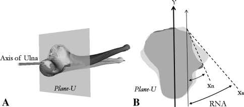
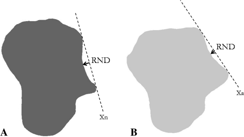
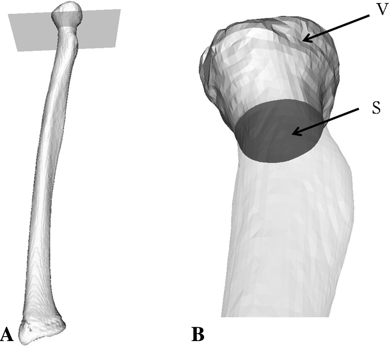
Patients and methods: We evaluated 15 patients with chronic radial head dislocations categorized by duration of "early" (< 3 years) (n = 8) and "longstanding" (> 3 years) (n = 7) groups. We measured the angle and depth of the radial notch of the proximal ulna and evaluated radial head deformity using 3-D bone models created from CT data.

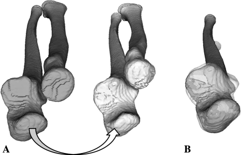
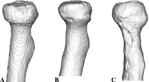
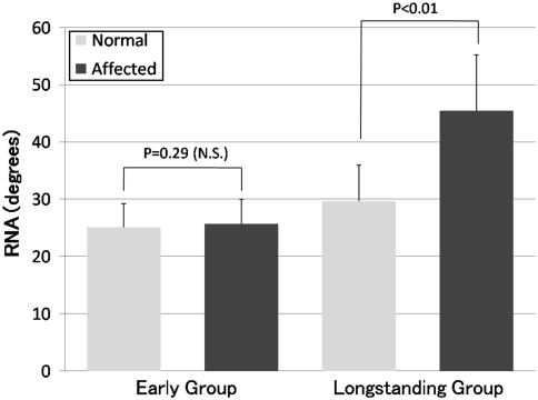
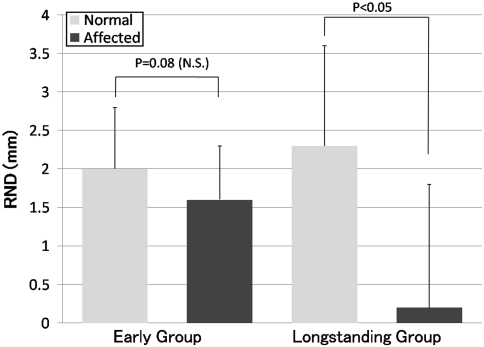
Results: For the early group, no differences were observed in the shape of the radial notch between affected and normal sides. For the longstanding group, the radial notch angle was greater on the affected side (mean +/- SD, 45.5 degrees +/- 9.7 degrees ) than on the normal side (29.7 degrees +/- 6.3 degrees ), and the radial notch depth was smaller on the affected side (0.2 +/- 1.6 mm) than on the normal side (2.3 +/- 1.3 mm). The shape of the radial head was nearly normal in the early group, whereas the longstanding group had a dome-shaped deformity.

Conclusions: In longstanding chronic radial head dislocation, deformation develops in the radial head and radial notch of the ulna, which is remodeled in a manner corresponding to the dislocated position of the radial head.

Level of evidence: Level III, prognostic study. See Guidelines for Authors for a complete description of levels of evidence.


Figures

Fig. 1A–B
Fig. 1A–B
(A) Mirror images of the 3-D bone models of the normal (gray) and affected (white) sides were created from CT data. (B) A mirror image of the normal ulna (gray) was superimposed on the affected ulna (translucent white).
Fig. 2A–B
Fig. 2A–B
(A) The plane perpendicular to the calculated axis of the ulna passing through the midpoint of the PRUJ is called “plane-U.” (B) The RNA is the angle between the line created by joining the ends of the radial notch (normal ulna: Line Xn, affected ulna: Line Xa) and the line passing through the ulnar posterior border and the tip of the coronoid process (Line Y).
Fig. 3A–B
Fig. 3A–B
The RND is the distance between Line X(n, a) and the bottom of the radial notch in (A) the contralateral normal ulna and (B) the affected ulna.
Fig. 4A–C
Fig. 4A–C
The patterns of the morphologic features of the radial head, (A) concave, (B) flat, and (C) domed, are shown.
Fig. 5A–B
Fig. 5A–B
(A) Plane-S is the plane at which the radial neck area is the smallest. (B) The minimal transverse area (S) and the volume of the radial head (V) are calculated.
Fig. 6
Fig. 6
No difference (p = 0.29) was observed in the RNA between the normal and affected sides in the early group. In the longstanding group, the RNA of the normal side was greater (p < 0.01) than that of the affected side because of an increase in the inclination of the radial notch. Bars = average; error bars = SD.
Fig. 7
Fig. 7
No difference (p = 0.08) was observed in the RND between the normal and affected sides in the early group. In the longstanding group, the RND of the affected side was smaller (p < 0.05) than that of the normal side owing to a shallower radial notch. Bar = average; error bars = SD.

Similar articles

Cited by

References

    1. Audette MA, Ferrie FP, Peters TM. An algorithmic overview of surface registration techniques for medical imaging. Med Image Anal. 2000;4:201–217. doi: 10.1016/S1361-8415(00)00014-1. - DOI - PubMed
    1. Austin R. Tardy palsy of the radial nerve from a Monteggia fracture. Injury. 1976;7:202–204. doi: 10.1016/0020-1383(76)90213-8. - DOI - PubMed
    1. Bado JL. The Monteggia lesion. Clin Orthop Relat Res. 1967;50:71–86. doi: 10.1097/00003086-196701000-00008. - DOI - PubMed
    1. Bell Tawse AJ. The treatment of malunited anterior Monteggia fractures in children. J Bone Joint Surg Br. 1965;47:718–723. - PubMed
    1. Besl PJ, Mackay N. A method for registration of 3-D shapes. IEEE Trans Patt Anal. 1992;14:239–256. doi: 10.1109/34.121791. - DOI

Publication types

MeSH terms