

How much are clinical fMRI reports influenced by standard postprocessing methods? An investigation of normalization and region of interest effects in the medial temporal lobe
- PMID: 20205247
- PMCID: PMC6870663
- DOI: 10.1002/hbm.20990
How much are clinical fMRI reports influenced by standard postprocessing methods? An investigation of normalization and region of interest effects in the medial temporal lobe
Abstract
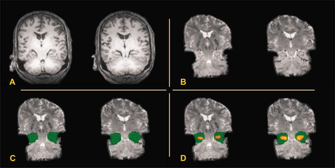
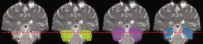
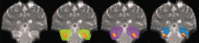
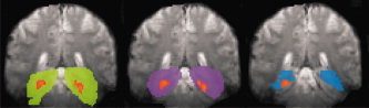
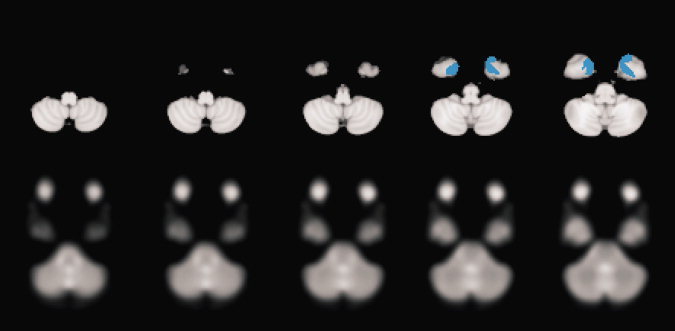
Recent evidence has indicated that standard postprocessing methods such as template-based region of interest (ROI) definition and normalization of individual brains to a standard template may influence final outcome of functional magnetic resonance imaging investigations. Here, we provide the first comprehensive investigation into whether ROI definition and normalization may also change the clinical interpretation of patient data. A series of medial temporal lobe epilepsy patients were investigated with a clinical memory paradigm and individually delineated as well as template-based ROIs. Different metrics for activation quantification were applied. Results show that the application of template-based ROIs can significantly change the clinical interpretation of individual patient data. This relates to sensitivity for brain activation and hemispheric dominance. We conclude that individual ROIs should be defined on nontransformed functional data and that use of more than one metric for activation quantification is beneficial.
Copyright © 2010 Wiley-Liss, Inc.
Figures
References
-
- Baxter L, Spencer B, Kerrigan JF ( 2007): Clinical application of functional MRI for memory using emotional enhancement: Deficit and recovery with limbic encephalitis. Epilepsy Behav 11: 454–459. - PubMed
-
- Beisteiner R, Erdler M, Teichtmeister C, Diemling M, Moser E, Edward V, Deecke L ( 1997): Magnetoencephalography may help to improve functional MRI brain mapping. Eur J Neurosci 9: 1072–1077. - PubMed
-
- Beisteiner R, Lanzenberger R, Novak K, Edward V, Windischberger C, Erdler M, Cunnington R, Gartus A, Streibl B, Moser E, Czech Th, Deecke L ( 2000): Improvement of presurgical patient evaluation by generation of functional magnetic resonance risk maps. Neurosci Lett 290: 13–16. - PubMed
-
- Beisteiner R, Windischberger C, Lanzenberger R, Edward V, Cunnington R, Erdler M, Gartus A, Streibl B, Moser E, Deecke L ( 2001): Finger somatotopy in human motor cortex. Neuroimage 13: 1016–1026. - PubMed
-
- Beisteiner R, Drabeck K, Foki T, Geissler A, Gartus A, Lehner‐Baumgartner E, Baumgartner C ( 2008): Does clinical memory fMRI provide a comprehensive map of medial temporal lobe structures? Experimental Neurology 213: 154–162. - PubMed
Publication types
MeSH terms
LinkOut - more resources
Full Text Sources
Medical
