. 2010 Mar 6:11:18.
doi: 10.1186/1471-2199-11-18.

DNA-PKcs plays a dominant role in the regulation of H2AX phosphorylation in response to DNA damage and cell cycle progression

Affiliations

DNA-PKcs plays a dominant role in the regulation of H2AX phosphorylation in response to DNA damage and cell cycle progression

Jing An et al. BMC Mol Biol. .

Abstract

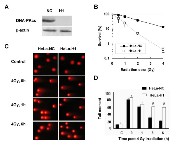
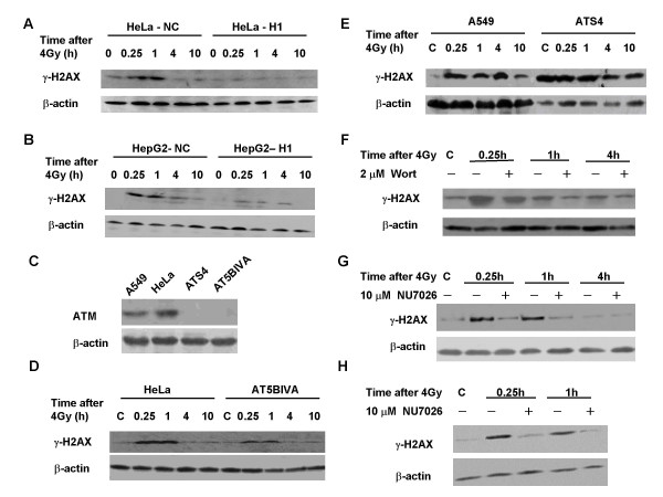
Background: When DNA double-strand breaks (DSB) are induced by ionizing radiation (IR) in cells, histone H2AX is quickly phosphorylated into gamma-H2AX (p-S139) around the DSB site. The necessity of DNA-PKcs in regulating the phosphorylation of H2AX in response to DNA damage and cell cycle progression was investigated.

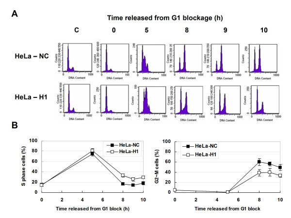
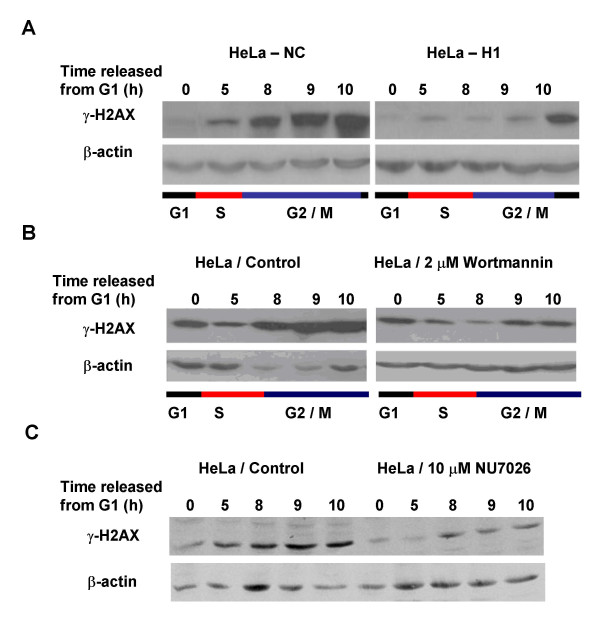
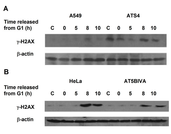
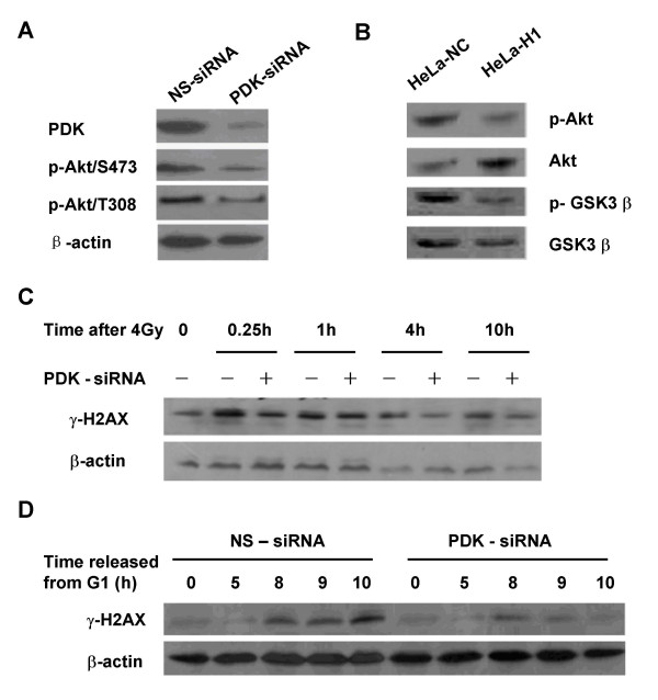
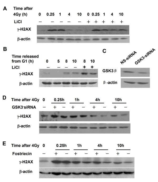
Results: The level of gamma H2AX in HeLa cells increased rapidly with a peak level at 0.25 - 1.0 h after 4 Gy gamma irradiation. SiRNA-mediated depression of DNA-PKcs resulted in a strikingly decreased level of gamma H2AX. An increased gamma H2AX was also induced in the ATM deficient cell line AT5BIVA at 0.5 - 1.0 h after 4 Gy gamma rays, and this IR-increased gamma H2AX in ATM deficient cells was dramatically abolished by the PIKK inhibitor wortmannin and the DNA-PKcs specific inhibitor NU7026. A high level of constitutive expression of gamma H2AX was observed in another ATM deficient cell line ATS4. The alteration of gamma H2AX level associated with cell cycle progression was also observed. HeLa cells with siRNA-depressed DNA-PKcs (HeLa-H1) or normal level DNA-PKcs (HeLa-NC) were synchronized at the G1 phase with the thymidine double-blocking method. At approximately 5 h after the synchronized cells were released from the G1 block, the S phase cells were dominant (80%) for both HeLa-H1 and HeLa-NC cells. At 8 - 9 h after the synchronized cells released from the G1 block, the proportion of G2/M population reached 56 - 60% for HeLa-NC cells, which was higher than that for HeLa H1 cells (33 - 40%). Consistently, the proportion of S phase for HeLa-NC cells decreased to approximately 15%; while a higher level (26 - 33%) was still maintained for the DNA-PKcs depleted HeLa-H1 cells during this period. In HeLa-NC cells, the gamma H2AX level increased gradually as the cells were released from the G1 block and entered the G2/M phase. However, this gamma H2AX alteration associated with cell cycle progressing was remarkably suppressed in the DNA-PKcs depleted HeLa-H1 cells, while wortmannin and NU7026 could also suppress this cell cycle related phosphorylation of H2AX. Furthermore, inhibition of GSK3 beta activity with LiCl or specific siRNA could up-regulate the gamma H2AX level and prolong the time of increased gamma H2AX to 10 h or more after 4 Gy. GSK3 beta is a negative regulation target of DNA-PKcs/Akt signaling via phosphorylation on Ser9, which leads to its inactivation. Depression of DNA-PKcs in HeLa cells leads to a decreased phosphorylation of Akt on Ser473 and its target GSK3 beta on Ser9, which, in other words, results in an increased activation of GSK3 beta. In addition, inhibition of PDK (another up-stream regulator of Akt/GSK3 beta) by siRNA can also decrease the induction of gamma H2AX in response to both DNA damage and cell cycle progression.

Conclusion: DNA-PKcs plays a dominant role in regulating the phosphorylation of H2AX in response to both DNA damage and cell cycle progression. It can directly phosphorylate H2AX independent of ATM and indirectly modulate the phosphorylation level of gamma H2AX via the Akt/GSK3 beta signal pathway.


Figures

Figure 1
Figure 1
Depletion of DNA-PKcs inhibits DNA double-strand break (DSB) repair and sensitizes HeLa cells to ionizing radiation. A: Depletion of DNA-PKcs by specific siRNA in HeLa-H1 cells. B: Survival curves of DNA-PKcs-depleted cells (HeLa-H1) and control cells (HeLa-NC). C: Comet images of DNA DSB detected by neutral single cell gel electrophoresis 0-6 h post-irradiation. D: The repair kinetics of 4 Gy-induced DNA DSBs detected by comet assay. The tail moment was used as the endpoint of DNA DSB. Each bar represents the mean tail moment from three independent experiments. 100 individual comets were counted per time point for each experiment. # p < 0.01, as compared to the HeLa-NC cells at the same time point.
Figure 2
Figure 2
Phosphorylation of H2AX in response to radiation-induced DNA damage in the presence or absence of DNA-PKcs and ATM. A, B: Phosphorylated H2AX (γH2AX) levels in DNA-PKcs depleted HeLa-H1 (A), HepG2-H1 [39] (B) cells and the control HeLa-NC or HepG2-NC cells. Cells were harvested at 0, 0.25, 1, 4 and 10 h after 4 Gy γ-irradiation. Protein expression was assayed by Western blotting. C: Expression of phospho-ATM at Ser-1981 detected at 0.5 h after 4 Gy γ-irradiation. D, E: H2AX phosphorylation in ATM-deficient cells AT5BIVA (D) and ATS4 (E) after 4 Gy γ-irradiation. Cells were harvested at 0, 0.25, 1, 4 and 10 h post-irradiation, and protein expression was assayed by Western blotting. F, G: The effect of the PI3K inhibitor wortmannin (F) and the DNA-PKcs specific inhibitor NU7026 (G) on H2AX phosphorylation in AT5BIVA cells after γ-irradiation. AT5BIVA cells were pretreated with 2 μM wortmannin or 10 μM NU7026 for 2 h, then irradiated with 4 Gy. The cells were harvested at 0, 0.25, 1 and 4 h after irradiation. Protein expression was assayed by Western blotting. H: The effect of NU7026 treatment (10 μM) on H2AX phosphorylation in HeLa-NC cells after 4 Gy irradiation.
Figure 3
Figure 3
Cell cycle progression of the synchronized DNA-PKcs-depressed HeLa-H1 cells and control HeLa-NC. A: Representative histograms of flow cytometry analysis. The cells synchronized in G1 phase by TdR double-blocking. G1 arrested cells were cultured in fresh DMEM, collected at 0, 5, 8, 9, 10 h after released from G1 block, analyzed by flow cytometry. B: Quantitative data of the cell cycle distribution of the cells released from G1 blockage by TdR double-blocking. Left panel, the proportion of S-phase cells at different times after released from G1 block. Right panel, the proportion of G2/M-phase cells at different times after cell cycle re-entry.
Figure 4
Figure 4
Phosphorylation of H2AX associated with cell cycle progression and the relative contributions of DNA-PKcs. A: Levels of γH2AX associated with cell cycle progression in DNA-PKcs-depleted HeLa-H1 (right) and control HeLa-NC cells (left). B: The effect of wortmannin on cell-cycle associated H2AX phosphorylation. After G1 synchronization by TdR double-blocking, cells were cultured in fresh DMEM supplemented with 2 μM wortmannin, then collected at 0, 5, 8, 9, 10 h, and analyzed by Western blotting. C: The effect of NU2076 on cell-cycle associated H2AX phosphorylation. After G1 synchronization by TdR double-blocking, cells were cultured in fresh DMEM supplemented with 10 μM NU2076, then collected at 0, 5, 8, 9, 10 h, and analyzed by Western blotting.
Figure 5
Figure 5
Cell Cycle associated H2AX phosphorylation in ATM deficient cells. The levels of phosphorylated H2AX at different times after release from G1 block were detected in ATM deficient cell lines ATS4 (A) and AT5BIVA (B). Synchronized G1 cells were cultured in fresh DMEM, harvested at 0, 5, 8 and 10 h after cell cycle re-entry, and protein expression was assayed by Western blotting.
Figure 6
Figure 6
Regulation of phosphoinositide-dependent kinase (PDK) on the phosphorylation of H2AX. A: RNAi mediated depletion of PDK protein. HeLa cells were transfected with 50 nM PDK specific siRNA molecules or non-specific (ns) control siRNA. Western blotting shows PDK expression. B: Phosphorylation of Akt at Ser473 and GSK3β at Ser9 was decreased in the DNA-PKcs depleted HeLa-H1 cells compared to control HeLa-NC cells. C: PDK regulates the phosphorylation of H2AX in response to DNA damage induced by 4 Gy of γ-irradiation. After 48 h incubation with 50 nM PDK-specific siRNA or non-specific (ns) control siRNA, cells were irradiated with 4 Gy γ rays, then harvested 0, 0.5, 1, 4, 10 h post-irradiation and analyzed by Western blotting. D: PDK regulates the phosphorylation of H2AX associated with cell cycle progression. After 24 h incubation with 50 nM PDK-specific siRNA or non-specific (ns) control siRNA, cells were synchronized in G1 phase by TdR double-blocking, then released and harvested after 5 h, for S-phase, and at 8, 9, and 10 h, for G2/M phase. The culture medium was supplemented with 50 nM siRNA molecules during the period of synchronization and cell cycle progression. Protein expression was assayed by Western blotting.
Figure 7
Figure 7
Regulation of H2AX phosphorylation in response to DNA damage and cell cycle progression by GSK3β. A: The GSK3β inhibitor LiCl prolongs phosphorylated H2AX increase in response to 4 Gy irradiation. HeLa cells were pretreated with 40 μM LiCl for 2 h, irradiated with 4 Gy γ rays and harvested at 0, 0.5, 1, 4 and 10 h after irradiation. Protein expression was assayed by Western blotting. B: GSK3β inhibitor LiCl enhanced the phosphorylation of H2AX in G2/M phase cells. To release the cells from G1 block and inhibit GSK3β activity, synchronized HeLa cells were grown in DMEM medium supplemented with 40 μM LiCl. S-phase cells were harvested at 5 h and G2/M phase cells at 8, 9 and10 h after released. Protein expression was assayed by Western blotting. C: RNAi depletion of GSK3β. HeLa cells were transfected with 50 nM GSK3β siRNA or non-specific (ns) control siRNA molecules. GSK3β expression was determined by Western blotting. D: Effect of GSK3β depletion on the phosphorylation of H2AX induced by 4 Gy γ-irradiation. After 48 h incubation with 50 nM GSK3β-specific siRNA or control non-specific (ns), cells were irradiated with 4 Gy γ rays, and harvested at 0, 0.25, 1, 4 and 10 h post-irradiation. Protein expression was assayed by Western blotting. E: Effect of the PP2A inhibitor fostriecin on H2AX phosphorylation. HeLa cells were pretreated with 50 nM fostriecin for 2 h, and then irradiated with 4 Gy γ rays, and harvested at 0, 0.25, 1, 4 and 10 h post-irradiation. Protein expression was assayed by Western blotting.

Similar articles

Cited by

References

    1. Albino AP, Huang X, Jorgensen ED, Gietl D, Traganos F, Darzynkiewicz Z. Induction of DNA double-strand breaks in A549 and normal human pulmonary epithelial cells by cigarette smoke is mediated by free radicals. Int J Oncol. 2006;28:1491–505. - PubMed
    1. Noel FL, Geraldine WLT. DNA repair: the importance of phosphorylating histone H2AX. Curr Biol. 2005;15:99–102. doi: 10.1016/j.cub.2005.01.029. - DOI - PubMed
    1. Rogakou EP, Pilch DR, Orr AH, Ivanova US, Bonner WM. DNA double-stranded breaks induce histone H2AX phophorylation on serine 139. J Biol Chem. 1998;273:5858–5868. doi: 10.1074/jbc.273.10.5858. - DOI - PubMed
    1. Celeste A, Petersen S, Romanienko PJ, Femandez-Capetillo O, Chen HT, Sedelnikova OA. Genomic instability in mice lacking histone H2AX. Science. 2002;296:922–927. doi: 10.1126/science.1069398. - DOI - PMC - PubMed
    1. Paull TT, Rogakou EP, Yamazaki V, Kirchgessner CU, Gellert M, Bonner WM. A critical role for histone H2AX in recruitment of repair factors to nuclear foci after DNA damage. Curr Biol. 2000;10:886–895. doi: 10.1016/S0960-9822(00)00610-2. - DOI - PubMed

Publication types

MeSH terms