

Review
doi: 10.1186/1746-1596-5-2.
Malignant peripheral nerve sheath tumor associated with neurofibromatosis type 1, with metastasis to the heart: a case report
Affiliations
- PMID: 20205747
- PMCID: PMC2881068
- DOI: 10.1186/1746-1596-5-2
Item in Clipboard
Review
Malignant peripheral nerve sheath tumor associated with neurofibromatosis type 1, with metastasis to the heart: a case report
Diagn Pathol.
.
Abstract
A rare case is presented of a 61-year-old man with a malignant peripheral nerve sheath tumor associated with neurofibromatosis type 1, with metastasis to the heart. The primary tumor originated in the right thigh in 1982. Since then, the patient has had repeated local recurrences in spite of repeated surgical treatment and adjuvant chemotherapy. He has developed previous metastases of the lung and heart. The patient died of cardiac involvement.
Figures
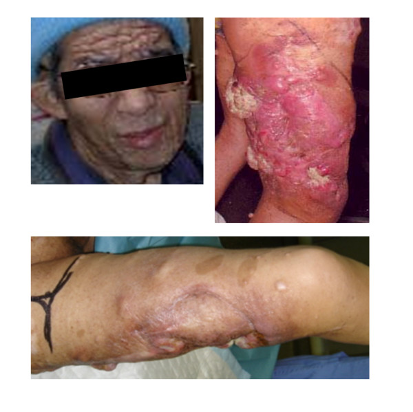
Clinical manifestations of NF1: multiple cutaneous neurofibromas and café-au-lait spots.
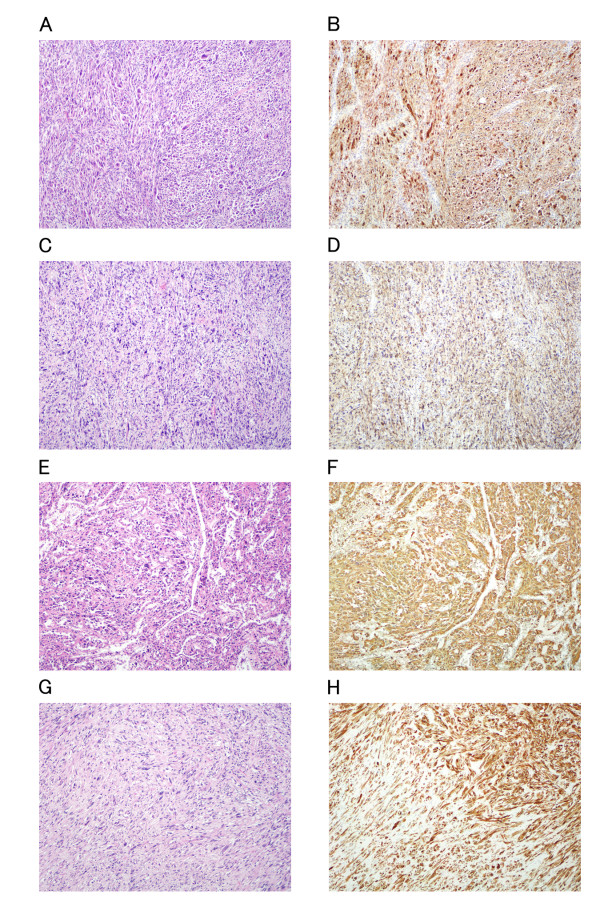
Hematoxylin and eosin and S-100 staining of the tumors of the thigh treated surgically in 1990 (A, B), recurrent tumor operated in 1995 (C, D) and metastatic tumor in heart (E, F) and lung (G, H) at autopsy in 2007. A, C, E, G: Hematoxylin and eosin staining; B, D, F, H: S-100 immunohistochemistry. Histological factors such as cellularity, degree of nuclear atypia, mitotic counts, and S-100 immunoreactivity were similar among the specimens.
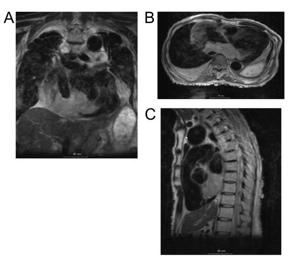
Magnetic resonance imaging showed a large tumor in the inferior-posterior wall of the heart. (A) Frontal cross section. Size: 7.5 cm. (B) Horizontal cross section. Size: 9 cm. (C) Sagittal cross section. Size: 6.5 cm.
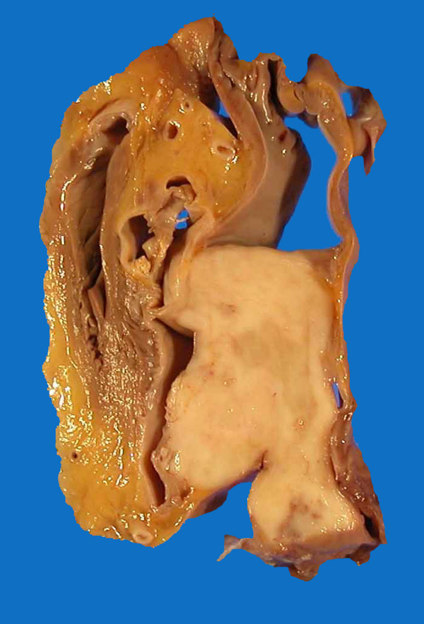
Autopsy specimen showing a tumor in the heart.
Similar articles
-
Long survival after resection for lung metastasis of malignant peripheral nerve sheath tumor in neurofibromatosis 1.Ann Thorac Cardiovasc Surg. 2008 Oct;14(5):322-4. Ann Thorac Cardiovasc Surg. 2008. PMID: 18989250
-
Spinal cord metastasis of a non-neurofibromatosis type-1 malignant peripheral nerve sheath tumor: an unusual manifestation of a rare tumor.J Neurooncol. 2005 Sep;74(2):183-5. doi: 10.1007/s11060-004-4596-4. J Neurooncol. 2005. PMID: 16193390
-
Osteosarcoma in a patient with neurofibromatosis type 1: a case report and review of the literature.Tohoku J Exp Med. 2006 Apr;208(4):343-8. doi: 10.1620/tjem.208.343. Tohoku J Exp Med. 2006. PMID: 16565597
-
Epithelioid malignant peripheral nerve sheath tumour: case report and review of the previously published cases.Cytopathology. 2002 Feb;13(1):54-63. doi: 10.1046/j.1365-2303.2002.00368.x. Cytopathology. 2002. PMID: 11985569 Review. No abstract available.
-
Fungating malignant peripheral nerve sheath tumor arising from a slow-growing mass in the forearm: a case report and review of the literature.J Med Case Rep. 2020 Jul 7;14(1):91. doi: 10.1186/s13256-020-02427-4. J Med Case Rep. 2020. PMID: 32631436 Free PMC article. Review.
Cited by
-
Ulnar malignant peripheral nerve sheath tumour diagnosis in a mixed-breed dog as a model to study human: histologic, immunohistochemical, and clinicopathologic study.Diagn Pathol. 2013 May 20;8:86. doi: 10.1186/1746-1596-8-86. Diagn Pathol. 2013. Retraction in: Diagn Pathol. 2016 Nov 2;11(1):121. doi: 10.1186/s13000-016-0570-7. PMID: 23688209 Free PMC article. Retracted.
-
Solitary neurofibroma of the gingiva with prominent differentiation of Meissner bodies: a case report.Diagn Pathol. 2010 Sep 22;5:61. doi: 10.1186/1746-1596-5-61. Diagn Pathol. 2010. PMID: 20858283 Free PMC article.
-
Propranolol Specifically Suppresses the Viability of Tumorous Schwann Cells Derived from Plexiform Neurofibromas In Vitro.In Vivo. 2020 May-Jun;34(3):1031-1036. doi: 10.21873/invivo.11872. In Vivo. 2020. PMID: 32354889 Free PMC article.
-
Malignant peripheral nerve sheath tumor of thigh with sphenoid and brain metastases: Extremely rare occurrence with dismal prognosis despite significant response to palliative chemoradiotherapy.World J Nucl Med. 2019 Jul-Sep;18(3):296-300. doi: 10.4103/wjnm.WJNM_54_18. World J Nucl Med. 2019. PMID: 31516375 Free PMC article.
-
Tumor targeted delivery of doxorubicin in malignant peripheral nerve sheath tumors.PLoS One. 2018 Jan 5;13(1):e0181529. doi: 10.1371/journal.pone.0181529. eCollection 2018. PLoS One. 2018. PMID: 29304038 Free PMC article.
References
-
- Weiss SW, Goldbrum JR. Enzinger and Weiss's Soft Tissue Tumors. St Louis: Mosby; 2008. Malignant tumors of peripheral nerves.
-
- Hussain R, Neligan MC. Metastatic malignant schwannoma in the heart. Ann Thorac Surg. 1993;56:374–375. - PubMed
-
- Menezes JAS, Greco OT, Fiorini M. Malignant schwannoma metastasizing to the heart. Arq Bras Cardiol. 1992;58:35–39. - PubMed
Publication types
MeSH terms
LinkOut - more resources
Full Text Sources
Research Materials
