. 2010 Jan 21:3:3.
doi: 10.1186/1756-6606-3-3.

DREAM (downstream regulatory element antagonist modulator) contributes to synaptic depression and contextual fear memory

Affiliations

DREAM (downstream regulatory element antagonist modulator) contributes to synaptic depression and contextual fear memory

Long-Jun Wu et al. Mol Brain. .

Abstract

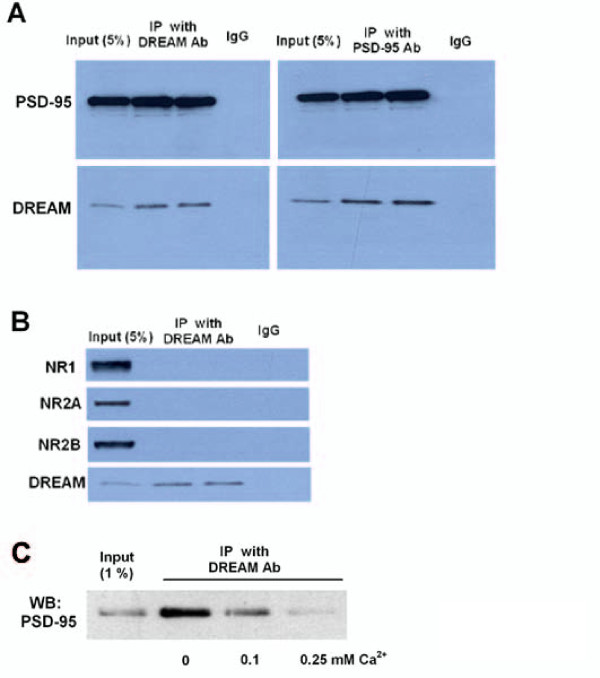
The downstream regulatory element antagonist modulator (DREAM), a multifunctional Ca2+-binding protein, binds specifically to DNA and several nucleoproteins regulating gene expression and with proteins outside the nucleus to regulate membrane excitability or calcium homeostasis. DREAM is highly expressed in the central nervous system including the hippocampus and cortex; however, the roles of DREAM in hippocampal synaptic transmission and plasticity have not been investigated. Taking advantage of transgenic mice overexpressing a Ca2+-insensitive DREAM mutant (TgDREAM), we used integrative methods including electrophysiology, biochemistry, immunostaining, and behavior tests to study the function of DREAM in synaptic transmission, long-term plasticity and fear memory in hippocampal CA1 region. We found that NMDA receptor but not AMPA receptor-mediated current was decreased in TgDREAM mice. Moreover, synaptic plasticity, such as long-term depression (LTD) but not long-term potentiation (LTP), was impaired in TgDREAM mice. Biochemical experiments found that DREAM interacts with PSD-95 and may inhibit NMDA receptor function through this interaction. Contextual fear memory was significantly impaired in TgDREAM mice. By contrast, sensory responses to noxious stimuli were not affected. Our results demonstrate that DREAM plays a novel role in postsynaptic modulation of the NMDA receptor, and contributes to synaptic plasticity and behavioral memory.


Figures

Figure 1
Figure 1
In vivo and in vitro characterization of TgDREAM. (A) Quantitative real-time RT-PCR of TgDREAM mRNA levels in different brain areas from transgenic mice. Expression level of the transgene was corrected by β-actin mRNA level in each ample. (B) Co-immunoprecipitation of TgDREAM-Flag and DREAM/KChIP Myc-tagged proteins (1 to 4) after overexpression in HEK293 cells.
Figure 2
Figure 2
Normal brain morphology in TgDREAM mice. Coronal sections showed no detectable morphological differences in the anterior cingulate cortex, somatosensory cortex, insular cortex, amygdala, hippocampus, thalamus, periaqueductal gray (PAG), spinal cord dorsal horn as well as dorsal root ganglia. Scale bar: 250 μm (cortex, amygdala and PAG), 500 μm (hippocampus and thalamus), 100 μm (spinal dorsal horn) and 50 μm (dorsal root ganglia).
Figure 3
Figure 3
Reduction of pairing protocol-induced long-term depression in the hippocampus in TgDREAM mice. (A) Diagram showing the paring protocol for LTP induction. The holding potential is +30 mV and 80 presynaptic stimulations at 2 Hz were applied. (B) LTP induced in hippocampal CA1 pyramidal neurons in wild-type mice (filled circles, n = 6) and TgDREAM mice (open circles, n = 6) by pairing protocol. In the present and following figures, the insets show averaged EPSC at 5 and 25 min after the pairing procedure (arrow). The dashed line indicates the mean basal synaptic response. (C) Pooled data show no significant difference (n.s.) of LTP in wild-type and TgDREAM mice. (D) Diagram showing the pairing protocol for LTD induction. The holding potential is -45 mV and 300 presynaptic stimulations at 1 Hz were applied. (E) LTD was stably induced by pairing protocol in wild-type mice (filled circles, n = 8), but completely abolished in TgDREAM mice (open circles, n = 8). (F) Pooled data show a significant difference in LTD between wild-type and TgDREAM mice. ** P < 0.01.
Figure 4
Figure 4
Reduced NMDA receptor-mediated synaptic transmission in TgDREAM mice. (A) Input-output relationship for NMDA receptor-mediated EPSCs evoked by various stimulation intensities in wild-type mice (filled circles, n = 8) and TgDREAM mice (open circles, n = 7). The amplitude of NMDA receptor current in TgDREAM mice was significantly reduced compared with that in wild-type mice. * P < 0.05. The right panel shows representative traces for NMDA receptor-mediated EPSCs in wild-type and TgDREAM mice. (B) The percentage of NR2A or NR2B component of NMDA receptor-mediated EPSCs is similar in the TgDREAM (n = 7) and wild-type mice (n = 7). The right panel shows sample traces of NMDAR-mediated EPSCs in control, 0.4 μM NVP-AAM077 (NVP) and 0.4 μM NVP-AAM077 + 3 μM ifenprodil (NVP + Ifen) in wild-type and TgDREAM mice.
Figure 5
Figure 5
Analysis of NMDA receptor protein in the hippocampus of TgDREAM mice. (A) Representative western blot (left) and quantified data (right) for expression levels of NR1, NR2A and NR2B subunits, and PSD-95 in hippocampus from wild-type and TgDREAM mice. Data were normalized to expression level of wild-type mice (n = 4 for each group). (B) Representative western blot (left) and quantified data (right) for phosphorylation of NR1, NR2A and NR2B at serine residues in hippocampus from wild-type and TgDREAM mice. Data were normalized to expression level of wild-type mice (n = 4 for each group).
Figure 6
Figure 6
DREAM interacts with PSD95 protein in vivo. (A) Co-immunoprecipitation of DREAM with PSD-95 in mouse hippocampal extracts from wild type mice. DREAM antibody immunoprecipitated DREAM bound to PSD-95 (left) while the PSD-95 antibody immunoprecipitated PSD-95 along with DREAM (right). Neither PSD-95 nor DREAM was immunoprecipitated using a control IgG. (B) DREAM antibody immunoprecipitated DREAM, but no NMDA receptor subunits (NR1, NR2A or NR2B) was detected in the immunoprecipitation. (C) PSD-95 protein was immunoprecipitated from mouse hippocampal extracts by a specific DREAM antibody. Increasing Ca2+ concentrations (0.1 and 0.25 mM) prevent this interaction. Absence of Ca2+ (0) corresponds to 2 mM EDTA.
Figure 7
Figure 7
Normal AMPA receptor function and expression inTgDREAM mice. (A) Input-output relationship for AMPA receptor-mediated EPSCs in wild-type (filled circles, n = 7) and TgDREAM mice (open circles, n = 7). There is no significant difference between the two groups. The sample traces are shown in the right panel. (B) Representative western blot (left) and quantified data (right) for expression levels of GluR1 and GluR2&3 subunits in hippocampus from wild-type and TgDREAM mice. Data were normalized to expression level of wild-type mice (n = 4 for each group). (C) Representative western blot (left) and quantified data (right) for phosphorylation of AMPA GluR1 receptor at ser831 and 845 sites in hippocampus from wild-type and TgDREAM mice. Data were normalized to expression level of wild-type mice (n = 4 for each group).
Figure 8
Figure 8
Impaired contextual fear memory in TgDREAM mice. (A) Contextual fear memory was impaired in TgDREAM mice (n = 8) compared with wild-type mice (n = 6) 1 h and 1 and 3 d after training. *** P < 0.001. (B) No significant difference in auditory fear conditioning between TgDREAM (n = 8) and wild-type mice (n = 6). (C) Comparison of nociceptive responses between TgDREAM (n = 8) and wild-type mice (n = 6). There was no significant difference in response latency between genotypes in the hotplate test (left) or in the tail-flick reflex (right).
Figure 9
Figure 9
Simplified model of the novel function of DREAM in NMDA receptor-mediated signaling transduction at synapses. At postsynaptic sites, DREAM interacts with PSD-95, and inhibits the functions of NMDA receptors. Binding of Ca2+ to DREAM releases the interaction between DREAM and PSD95. In TgDREAM mice, Ca2+-insensitive mutant DREAM may constitutively bind to PSD95, leading to the impaired recruitment of PSD-95 and reduced NMDA receptor function. The loss of LTD in TgDREAM mice is likely due to the impaired function of NMDA receptors. Within the nucleus, it is known that DREAM binds to DRE located downstream from the transcriptional start site, and inhibits the promoter activity. The binding of Ca2+ to DREAM releases this inhibition, leading to higher levels of transcription.

Similar articles

Cited by

References

    1. Hyman SE, Malenka RC. Addiction and the brain: the neurobiology of compulsion and its persistence. Nat Rev Neurosci. 2001;2:695–703. doi: 10.1038/35094560. - DOI - PubMed
    1. Kandel ER. The molecular biology of memory storage: a dialogue between genes and synapses. Science. 2001;294:1030–1038. doi: 10.1126/science.1067020. - DOI - PubMed
    1. Nestler EJ. Molecular basis of long-term plasticity underlying addiction. Nat Rev Neurosci. 2001;2:119–128. doi: 10.1038/35053570. - DOI - PubMed
    1. Zhuo M. Neuronal mechanism for neuropathic pain. Mol Pain. 2007;3:14. doi: 10.1186/1744-8069-3-14. - DOI - PMC - PubMed
    1. Woolf CJ, Salter MW. Neuronal plasticity: increasing the gain in pain. Science. 2000;288:1765–1769. doi: 10.1126/science.288.5472.1765. - DOI - PubMed

Publication types

Substances