

Role of senescence and mitotic catastrophe in cancer therapy
- PMID: 20205872
- PMCID: PMC2827387
- DOI: 10.1186/1747-1028-5-4
Role of senescence and mitotic catastrophe in cancer therapy
Retraction in
-
Retraction: Role of senescence and mitotic catastrophe in cancer therapy.Cell Div. 2012 Jul 12;7:15. doi: 10.1186/1747-1028-7-15. eCollection 2012. Cell Div. 2012. PMID: 22587525 Free PMC article. No abstract available.
Abstract
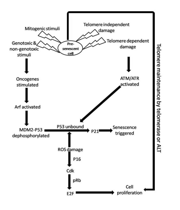
Senescence and mitotic catastrophe (MC) are two distinct crucial non-apoptotic mechanisms, often triggered in cancer cells and tissues in response to anti-cancer drugs. Chemotherapeuticals and myriad other factors induce cell eradication via these routes. While senescence drives the cells to a state of quiescence, MC drives the cells towards death during the course of mitosis. The senescent phenotype distinguishes tumor cells that survived drug exposure but lost the ability to form colonies from those that recover and proliferate after treatment. Although senescent cells do not proliferate, they are metabolically active and may secrete proteins with potential tumor-promoting activities. The other anti-proliferative response of tumor cells is MC that is a form of cell death that results from abnormal mitosis and leads to the formation of interphase cells with multiple micronuclei. Different classes of cytotoxic agents induce MC, but the pathways of abnormal mitosis differ depending on the nature of the inducer and the status of cell-cycle checkpoints. In this review, we compare the two pathways and mention that they are activated to curb the growth of tumors. Altogether, we have highlighted the possibilities of the use of senescence targeting drugs, mitotic kinases and anti-mitotic agents in fabricating novel strategies in cancer control.
Figures

Similar articles
-
If not apoptosis, then what? Treatment-induced senescence and mitotic catastrophe in tumor cells.Drug Resist Updat. 2001 Oct;4(5):303-13. doi: 10.1054/drup.2001.0213. Drug Resist Updat. 2001. PMID: 11991684 Review.
-
Mitotic catastrophe as a consequence of chemotherapy.Anticancer Agents Med Chem. 2006 Nov;6(6):589-602. doi: 10.2174/187152006778699086. Anticancer Agents Med Chem. 2006. PMID: 17100562 Review.
-
Clonogenic Assays to Detect Cell Fate in Mitotic Catastrophe.Methods Mol Biol. 2021;2267:227-239. doi: 10.1007/978-1-0716-1217-0_16. Methods Mol Biol. 2021. PMID: 33786796
-
Mechanisms of drug-induced mitotic catastrophe in cancer cells.Curr Pharm Des. 2010 Jan;16(1):69-78. doi: 10.2174/138161210789941801. Curr Pharm Des. 2010. PMID: 20214619 Review.
-
Two distinct modes of cell death induced by doxorubicin: apoptosis and cell death through mitotic catastrophe accompanied by senescence-like phenotype.Oncogene. 2005 Jul 14;24(30):4765-77. doi: 10.1038/sj.onc.1208627. Oncogene. 2005. PMID: 15870702
Cited by
-
Silencing erythropoietin receptor on glioma cells reinforces efficacy of temozolomide and X-rays through senescence and mitotic catastrophe.Oncotarget. 2015 Feb 10;6(4):2101-19. doi: 10.18632/oncotarget.2937. Oncotarget. 2015. PMID: 25544764 Free PMC article.
-
A radiosensitizer, gallotannin-rich extract from Bouea macrophylla seeds, inhibits radiation-induced epithelial-mesenchymal transition in breast cancer cells.BMC Complement Med Ther. 2021 Jul 3;21(1):189. doi: 10.1186/s12906-021-03363-6. BMC Complement Med Ther. 2021. PMID: 34217266 Free PMC article.
-
Glioma pathogenesis-related protein 1 induces prostate cancer cell death through Hsc70-mediated suppression of AURKA and TPX2.Mol Oncol. 2013 Jun;7(3):484-96. doi: 10.1016/j.molonc.2012.12.005. Epub 2012 Dec 31. Mol Oncol. 2013. PMID: 23333597 Free PMC article.
-
Radiation-induced cellular and molecular alterations in asexual intraerythrocytic Plasmodium falciparum.J Infect Dis. 2013 Jan 1;207(1):164-74. doi: 10.1093/infdis/jis645. Epub 2012 Oct 24. J Infect Dis. 2013. PMID: 23100570 Free PMC article.
-
Retraction: Role of senescence and mitotic catastrophe in cancer therapy.Cell Div. 2012 Jul 12;7:15. doi: 10.1186/1747-1028-7-15. eCollection 2012. Cell Div. 2012. PMID: 22587525 Free PMC article. No abstract available.
References
Publication types
LinkOut - more resources
Full Text Sources
