

Cigarette smoke regulates VEGFR2-mediated survival signaling in rat lungs
- PMID: 20205917
- PMCID: PMC2831890
- DOI: 10.1186/1476-9255-7-11
Cigarette smoke regulates VEGFR2-mediated survival signaling in rat lungs
Abstract
Background: Vascular endothelial growth factor (VEGF) and VEGF receptor 2 (VEGFR2)-mediated survival signaling is critical to endothelial cell survival, maintenance of the vasculature and alveolar structure and regeneration of lung tissue. Reduced VEGF and VEGFR2 expression in emphysematous lungs has been linked to increased endothelial cell death and vascular regression. Previously, we have shown that CS down-regulated the VEGFR2 and its downstream signaling in mouse lungs. However, the VEGFR2-mediated survival signaling in response to oxidants/cigarette smoke (CS) is not known. We hypothesized that CS exposure leads to disruption of VEGFR2-mediated endothelial survival signaling in rat lungs.
Methods: Adult male Sprague-Dawley rats were exposed CS for 3 days, 8 weeks and 6 months to investigate the effect of CS on VEGFR2-mediated survival signaling by measuring the Akt/PI3-kinase/eNOS downstream signaling in rat lungs.
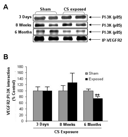
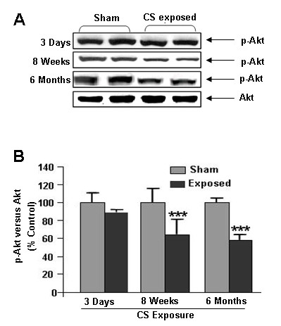
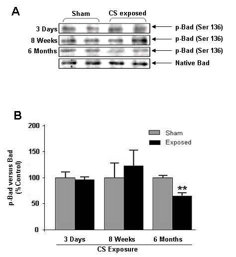
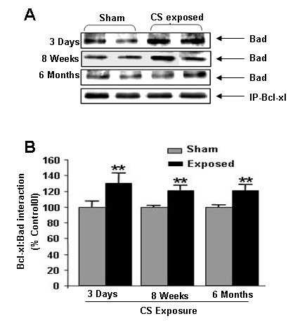
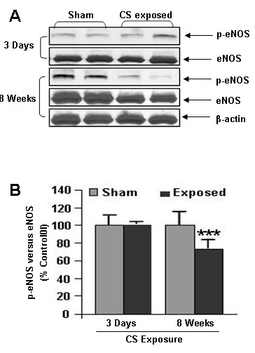
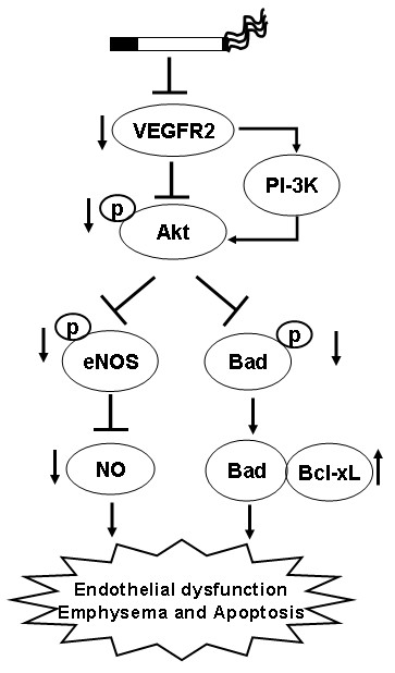
Results and discussion: We show that CS disrupts VEGFR2/PI3-kinase association leading to decreased Akt and eNOS phosphorylation. This may further alter the phosphorylation of the pro-apoptotic protein Bad and increase the Bad/Bcl-xl association. However, this was not associated with a significant lung cell death as evidenced by active caspase-3 levels. These data suggest that although CS altered the VEGFR2-mediated survival signaling in the rat lungs, but it was not sufficient to cause lung cell death.
Conclusion: The rat lungs exposed to CS in acute, sub-chronic and chronic levels may be representative of smokers where survival signaling is altered but was not associated with lung cell death whereas emphysema is known to be associated with lung cell apoptosis.
Figures


Similar articles
-
Cigarette-smoke-induced oxidative/nitrosative stress impairs VEGF- and fluid-shear-stress-mediated signaling in endothelial cells.Antioxid Redox Signal. 2010 Jun 15;12(12):1355-69. doi: 10.1089/ars.2009.2874. Antioxid Redox Signal. 2010. Retraction in: Antioxid Redox Signal. 2013 Apr 20;18(12):1535. doi: 10.1089/ars.2012.5170. PMID: 19929443 Free PMC article. Retracted.
-
Cigarette smoke-mediated oxidative stress, shear stress, and endothelial dysfunction: role of VEGFR2.Ann N Y Acad Sci. 2010 Aug;1203:66-72. doi: 10.1111/j.1749-6632.2010.05601.x. Ann N Y Acad Sci. 2010. PMID: 20716285 Review.
-
Cigarette smoke disrupts VEGF165-VEGFR-2 receptor signaling complex in rat lungs and patients with COPD: morphological impact of VEGFR-2 inhibition.Am J Physiol Lung Cell Mol Physiol. 2006 May;290(5):L897-908. doi: 10.1152/ajplung.00116.2005. Epub 2005 Dec 16. Am J Physiol Lung Cell Mol Physiol. 2006. PMID: 16361360
-
Glomerular endothelial PI3 kinase-α couples to VEGFR2, but is not required for eNOS activation.Am J Physiol Renal Physiol. 2011 Dec;301(6):F1242-50. doi: 10.1152/ajprenal.00662.2010. Epub 2011 Sep 21. Am J Physiol Renal Physiol. 2011. PMID: 21937609
-
Cigarette smoke-induced lung endothelial apoptosis and emphysema are associated with impairment of FAK and eIF2α.Microvasc Res. 2014 Jul;94:80-9. doi: 10.1016/j.mvr.2014.05.003. Epub 2014 May 20. Microvasc Res. 2014. PMID: 24853558 Free PMC article.
Cited by
-
Dopamine inhibits pulmonary edema through the VEGF-VEGFR2 axis in a murine model of acute lung injury.Am J Physiol Lung Cell Mol Physiol. 2012 Jan 15;302(2):L185-92. doi: 10.1152/ajplung.00274.2010. Epub 2011 Oct 14. Am J Physiol Lung Cell Mol Physiol. 2012. PMID: 22003095 Free PMC article.
-
The Role of Mitochondria and Oxidative/Antioxidative Imbalance in Pathobiology of Chronic Obstructive Pulmonary Disease.Oxid Med Cell Longev. 2016;2016:7808576. doi: 10.1155/2016/7808576. Epub 2016 Dec 26. Oxid Med Cell Longev. 2016. PMID: 28105251 Free PMC article. Review.
-
Gender differences in plasma biomarker levels in a cohort of COPD patients: a pilot study.PLoS One. 2011 Jan 18;6(1):e16021. doi: 10.1371/journal.pone.0016021. PLoS One. 2011. PMID: 21267454 Free PMC article.
-
Acrolein inhalation prevents vascular endothelial growth factor-induced mobilization of Flk-1+/Sca-1+ cells in mice.Arterioscler Thromb Vasc Biol. 2011 Jul;31(7):1598-606. doi: 10.1161/ATVBAHA.111.227124. Epub 2011 Apr 28. Arterioscler Thromb Vasc Biol. 2011. PMID: 21527748 Free PMC article.
-
Tobacco smoke induced COPD/emphysema in the animal model-are we all on the same page?Front Physiol. 2013 May 15;4:91. doi: 10.3389/fphys.2013.00091. eCollection 2013. Front Physiol. 2013. PMID: 23720629 Free PMC article.
References
-
- Kim WD, Eidelman DH, Izquierdo JL, Ghezzo H, Saetta MP, Cosio MG. Centrilobular and panlobular emphysema in smokers. Two distinct morphologic and functional entities. Am Rev Respir Dis. 1991;144:1385–1390. - PubMed
-
- Kasahara Y, Tuder RM, Cool CD, Lynch DA, Flores SC, Voelkel NF. Endothelial cell death and decreased expression of vascular endothelial growth factor and vascular endothelial growth factor receptor 2 in emphysema. Am J Respir Crit Care Med. 2001;163:737–744. - PubMed
Grants and funding
LinkOut - more resources
Full Text Sources
Research Materials
