

Pancratistatin induces apoptosis in clinical leukemia samples with minimal effect on non-cancerous peripheral blood mononuclear cells
- PMID: 20205924
- PMCID: PMC2845577
- DOI: 10.1186/1475-2867-10-6
Pancratistatin induces apoptosis in clinical leukemia samples with minimal effect on non-cancerous peripheral blood mononuclear cells
Abstract
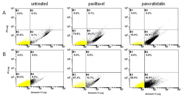
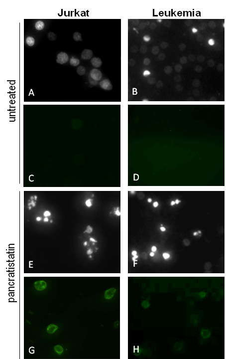
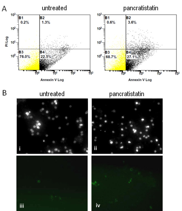
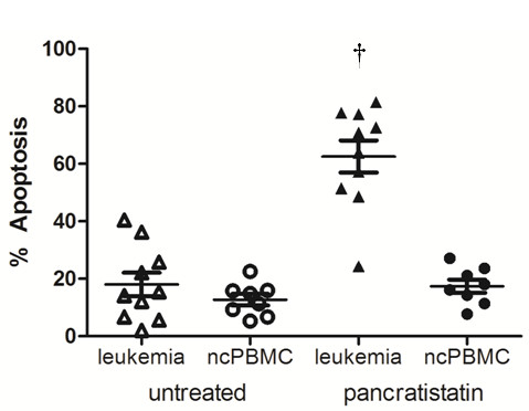
Background: Pancratistatin, a natural compound extracted from Hymenocallis littoralis, can selectively induce apoptosis in several cancer cell lines. In this ex vivo study, we evaluated the effect of pancratistatin on peripheral blood mononuclear cells obtained from 15 leukemia patients prior to clinical intervention of newly diagnosed patients, as well as others of different ages in relapse and at various disease progression states.
Results: Mononuclear cells from healthy volunteers and leukemia patients were exposed to 1 microM pancratistatin for up to 48 h. Irrespective of leukemia type, pancratistatin induced apoptosis in the leukemic samples, with minimal effects on non-cancerous peripheral blood mononuclear control cells.
Conclusion: Our results show that pancratistatin is an effective and selective anti-cancer agent with potential for advancement to clinical trials.
Figures
Similar articles
-
Induction of apoptotic cell death specifically in rat and human cancer cells by pancratistatin.Artif Cells Blood Substit Immobil Biotechnol. 2005;33(3):279-95. doi: 10.1081/bio-200066621. Artif Cells Blood Substit Immobil Biotechnol. 2005. PMID: 16152693
-
Pancratistatin induces apoptosis and autophagy in metastatic prostate cancer cells.Int J Oncol. 2011 Jun;38(6):1549-56. doi: 10.3892/ijo.2011.977. Epub 2011 Mar 17. Int J Oncol. 2011. PMID: 21424119
-
Pancratistatin causes early activation of caspase-3 and the flipping of phosphatidyl serine followed by rapid apoptosis specifically in human lymphoma cells.Cancer Chemother Pharmacol. 2005 Jul;56(1):29-38. doi: 10.1007/s00280-004-0941-8. Epub 2005 Feb 22. Cancer Chemother Pharmacol. 2005. PMID: 15726366
-
Antineoplastic agents, 294. Variations in the formation of pancratistatin and related isocarbostyrils in Hymenocallis littoralis.J Nat Prod. 1995 Jan;58(1):37-43. doi: 10.1021/np50115a004. J Nat Prod. 1995. PMID: 7760076
-
Sensitization of human melanoma cells by tamoxifen to apoptosis induction by pancratistatin, a nongenotoxic natural compound.Melanoma Res. 2011 Feb;21(1):1-11. doi: 10.1097/CMR.0b013e328337abff. Melanoma Res. 2011. PMID: 20300039
Cited by
-
Incredible use of plant-derived bioactives as anticancer agents.RSC Adv. 2025 Jan 20;15(3):1721-1746. doi: 10.1039/d4ra05089d. eCollection 2025 Jan 16. RSC Adv. 2025. PMID: 39835210 Free PMC article. Review.
-
ZYH005, a novel DNA intercalator, overcomes all-trans retinoic acid resistance in acute promyelocytic leukemia.Nucleic Acids Res. 2018 Apr 20;46(7):3284-3297. doi: 10.1093/nar/gky202. Nucleic Acids Res. 2018. PMID: 29554366 Free PMC article.
-
Total Synthesis of (+)-Pancratistatin by the Rh(III)-Catalyzed Addition of a Densely Functionalized Benzamide to a Sugar-Derived Nitroalkene.Org Lett. 2017 Jun 2;19(11):2985-2988. doi: 10.1021/acs.orglett.7b01220. Epub 2017 May 24. Org Lett. 2017. PMID: 28537406 Free PMC article.
-
Discovery of new anticancer agents from higher plants.Front Biosci (Schol Ed). 2012 Jan 1;4(1):142-56. doi: 10.2741/s257. Front Biosci (Schol Ed). 2012. PMID: 22202049 Free PMC article. Review.
-
Enhancement of apoptotic and autophagic induction by a novel synthetic C-1 analogue of 7-deoxypancratistatin in human breast adenocarcinoma and neuroblastoma cells with tamoxifen.J Vis Exp. 2012 May 30;(63):3586. doi: 10.3791/3586. J Vis Exp. 2012. PMID: 22688195 Free PMC article.
References
-
- Siedlakowski P, McLachlan-Burgess A, Griffin C, Tirumalai SS, McNulty J, Pandey S. Synergy of Pancratistatin and Tamoxifen on breast cancer cells in inducing apoptosis by targeting mitochondria. Cancer Biol Ther. 2007;7(3):376–384. - PubMed
-
- Dumont P, Ingrassia L, Rouzeau S, Ribaucour F, Thomas S, Roland I, Darro F, Lefranc F, Kiss R. The Amaryllidaceae isocarbostyril narciclasine induces apoptosis by activation of the death receptor and/or mitochondrial pathways in cancer cells but not in normal fibroblasts. Neoplasia. 2007;9(9):766–76. doi: 10.1593/neo.07535. - DOI - PMC - PubMed
-
- Gerber DE. Targeted therapies: a new generation of cancer treatments. Amer Fam Physician. 2008;77(3):311–319. - PubMed
LinkOut - more resources
Full Text Sources
