. 2010 Mar 8;11(1):28.
doi: 10.1186/1465-9921-11-28.

Elevation of IL-6 in the allergic asthmatic airway is independent of inflammation but associates with loss of central airway function

Affiliations

Elevation of IL-6 in the allergic asthmatic airway is independent of inflammation but associates with loss of central airway function

Wendy A Neveu et al. Respir Res. .

Abstract

Background: Asthma is a chronic inflammatory disease of the airway that is characterized by a Th2-type of immune response with increasing evidence for involvement of Th17 cells. The role of IL-6 in promoting effector T cell subsets suggest that IL-6 may play a functional role in asthma. Classically IL-6 has been viewed as an inflammatory marker, along with TNFalpha and IL-1beta, rather than as regulatory cytokine.

Objective: To investigate the potential relationship between IL-6 and other proinflammatory cytokines, Th2/Th17 cytokines and lung function in allergic asthma, and thus evaluate the potential role of IL-6 in this disease.

Methods: Cytokine levels in induced sputum and lung function were measured in 16 healthy control and 18 mild-moderate allergic asthmatic subjects.

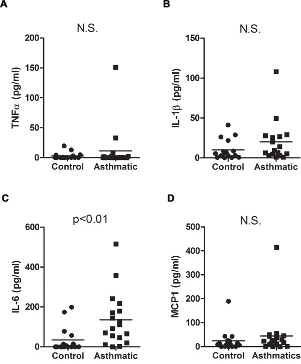
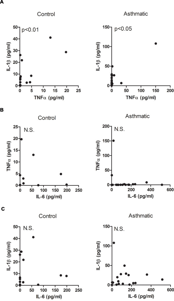
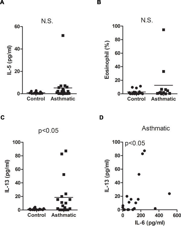
Results: The levels of the proinflammatory biomarkers TNFalpha and IL-1beta were not different between the control and asthmatic group. In contrast, IL-6 levels were specifically elevated in asthmatic subjects compared with healthy controls (p < 0.01). Hierarchical regression analysis in the total study cohort indicates that the relationship between asthma and lung function could be mediated by IL-6. Among Th2 cytokines only IL-13 (p < 0.05) was also elevated in the asthmatic group, and positively correlated with IL-6 levels (rS = 0.53, p < 0.05).

Conclusions: In mild-moderate asthma, IL-6 dissociates from other proinflammatory biomarkers, but correlates with IL-13 levels. Furthermore, IL-6 may contribute to impaired lung function in allergic asthma.


Figures

Figure 1
Figure 1
IL-6 but not TNFα or IL-1β is elevated in induced sputum of mild-moderate asthmatic subjects. (A) TNFα, (B) IL-1β, (C) IL-6 and (D) MCP-1 levels were analyzed in induced sputum of mild-moderate asthmatic subjects (n = 18) and healthy controls (n = 16).
Figure 2
Figure 2
While TNFα correlates with IL-1β in induced sputum, neither correlates with IL-6 in mild-moderate asthmatic subjects. (A) Correlative analysis of TNFα and IL-1β levels in control (rs = 0.64, p < 0.01) and asthmatic subjects (rs = 0.49, p < 0.05). (B) Correlative analysis of IL-6 and TNFα levels in control (rs = 0.46, p = 0.08) and asthmatic subjects (rs = 0.29, p = 0.24). (C) Correlative analysis of IL-6 and IL-1β levels in control (rs = 0.38, p = 0.15) and asthmatic subjects (rs = 0.18, p = 0.48).
Figure 3
Figure 3
Relationship of IL-6 with lung function in mild-moderate asthmatic subjects. (A) Correlative analysis of sputum IL-6 levels and FEV1/FVC ratio in control (rs = -0.18, p = 0.50) and asthmatic (rs = -0.23, p = 0.37) subjects. (B) Correlative analysis of sputum IL-6 levels with PEFR in control (rs = -0.26, p = 0.34) and asthmatic (rs = -0.37, p = 0.13) subjects. (C) Schematic representation of the hierarchical regression analysis in the total cohort. Path X is the correlation between IL-6 levels and disease status (asthma). Path Y is the correlation between IL-6 levels and lung function. Path Z is the correlation between disease status and lung function.
Figure 4
Figure 4
IL-17 levels in mild-moderate asthmatic subjects and its relationship to IL-6. (A) IL-17 levels were analyzed in control and asthmatic subjects. (B) Correlative analysis of IL-17 levels with percentage of neutrophils in asthmatic subjects (rs = 0.60, p < 0.05). (C) Correlative analysis of IL-6 levels with percentage of neutrophils in asthmatic subjects (rs = 0.07, p = 0.83). (D) Correlative analysis of IL-6 and IL-17 levels in asthmatic subjects (rs = -0.45, p = 0.06).
Figure 5
Figure 5
Selective elevation of IL-13 in mild-moderate asthmatic subjects compared with healthy control subjects. (A) IL-5 levels and (B) percentage of eosinophils in induced sputum were analyzed in the control and asthmatic groups. (C) IL-13 levels were analyzed in control and asthmatic subjects. (D) Correlative analysis of IL-6 and IL-13 levels in the asthmatic group (rs = 0.53, p < 0.05).

Similar articles

Cited by

References

    1. Georas SN, Guo J, De Fanis U, Casolaro V. T-helper cell type-2 regulation in allergic disease. Eur Respir J. 2005;26(6):1119–1137. doi: 10.1183/09031936.05.00006005. - DOI - PubMed
    1. Schnyder-Candrian S, Togbe D, Couillin I, Mercier I, Brombacher F, Quesniaux V, Fossiez F, Ryffel B, Schnyder B. Interleukin-17 is a negative regulator of established allergic asthma. The Journal of experimental medicine. 2006;203(12):2715–2725. doi: 10.1084/jem.20061401. - DOI - PMC - PubMed
    1. Ouyang W, Kolls JK, Zheng Y. The biological functions of T helper 17 cell effector cytokines in inflammation. Immunity. 2008;28(4):454–467. doi: 10.1016/j.immuni.2008.03.004. - DOI - PMC - PubMed
    1. Al-Ramli W, Al Samri M, Hamid Q. TH-17 cell-related cytokines' potential role in the pathogenesis of severe asthma. J Asthma. 2008;45(S1):41–44. doi: 10.1080/02770900802594759. - DOI - PubMed
    1. Cromwell O, Hamid Q, Corrigan CJ, Barkans J, Meng Q, Collins PD, Kay AB. Expression and generation of interleukin-8, IL-6 and granulocyte-macrophage colony-stimulating factor by bronchial epithelial cells and enhancement by IL-1 beta and tumour necrosis factor-alpha. Immunology. 1992;77(3):330–337. - PMC - PubMed

Publication types