

Functional significance of the hepaCAM gene in bladder cancer
- PMID: 20205955
- PMCID: PMC2845116
- DOI: 10.1186/1471-2407-10-83
Functional significance of the hepaCAM gene in bladder cancer
Abstract
Background: The hepaCAM gene encodes a new immunoglobulin-like cell adhesion molecule, and its expression is suppressed in a variety of human cancers. Additionally, hepaCAM possesses properties often observed in tumor suppressor genes. However, the expression and biological function of hepaCAM has not been investigated in bladder cancer. Therefore we sought to examine hepaCAM expression and the relationship between its structure and function in human transitional cell carcinoma of bladder (TCCB).
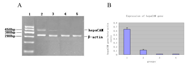
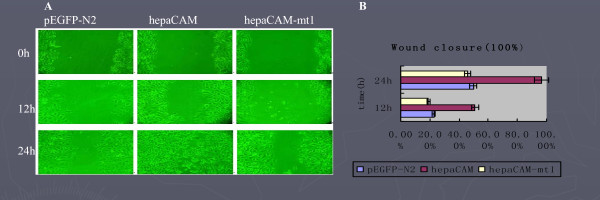
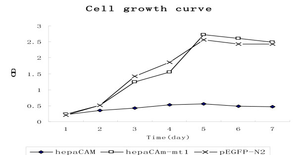
Materials and methods: HepaCAM expression was evaluated in 28 normal and 34 TCCB bladder specimens and 2 TCCB cell lines using semi-quantitative RT-PCR. The wild-type hepaCAM and the extracellular domain-truncated mutant gene were transfected into the TCCB cell line T24, and the biological properties of both the wild-type gene and the domain-truncated mutant were then assessed.
Results: HepaCAM expression was down-regulated in 82% (28/34) of TCCB specimens and undetectable in the 2 TCCB cell lines tested. The localization of hepaCAM appeared to be dependent on cell density in T24 cells. In widely spread cells, hepaCAM accumulated on the perinuclear membrane and the cell surface protrusions, whereas in confluent cells, hepaCAM was predominantly localized at the sites of cell-cell contacts on the cell membrane. Functionally, hepaCAM expressed not only increased cell spreading, delayed cell detachment, enhanced wound healing and increased cell invasion; it also inhibited cell growth (P < 0.01). When the extracellular domain was deleted, the localization of hepaCAM was significantly altered, and it lost both its adhesive function and its influence on cell growth.
Conclusions: HepaCAM is involved in cell adhesion and growth control, and its expression is frequently silenced in TCCB. The extracellular domain of hepaCAM is essential to its physiological and biological functions.
Figures

Similar articles
-
hepaCAM and p-mTOR closely correlate in bladder transitional cell carcinoma and hepaCAM expression inhibits proliferation via an AMPK/mTOR dependent pathway in human bladder cancer cells.J Urol. 2013 Nov;190(5):1912-8. doi: 10.1016/j.juro.2013.05.013. Epub 2013 May 10. J Urol. 2013. PMID: 23669565
-
[Construction of deltaNp63 specific small hairpin RNA expressing plasmid and its role in bladder cancer--a preliminary study].Zhonghua Zhong Liu Za Zhi. 2006 Nov;28(11):820-5. Zhonghua Zhong Liu Za Zhi. 2006. PMID: 17416002 Chinese.
-
Cloning and characterization of hepaCAM, a novel Ig-like cell adhesion molecule suppressed in human hepatocellular carcinoma.J Hepatol. 2005 Jun;42(6):833-41. doi: 10.1016/j.jhep.2005.01.025. Epub 2005 Apr 7. J Hepatol. 2005. PMID: 15885354
-
Exploration of the correlations between interferon-γ in patient serum and HEPACAM in bladder transitional cell carcinoma, and the interferon-γ mechanism inhibiting BIU-87 proliferation.J Urol. 2012 Oct;188(4):1346-53. doi: 10.1016/j.juro.2012.06.005. Epub 2012 Aug 17. J Urol. 2012. PMID: 22906662
-
[Influence of hepatocyte cell adhesion molecule on gene expression profile of human bladder transitional cell carcinoma cell line].Zhongguo Yi Xue Ke Xue Yuan Xue Bao. 2013 Apr;35(2):190-8. doi: 10.3881/j.issn.1000-503X.2013.02.012. Zhongguo Yi Xue Ke Xue Yuan Xue Bao. 2013. PMID: 23643009 Chinese.
Cited by
-
Frequent alterations of LOH11CR2A, PIG8 and CHEK1 genes at chromosomal 11q24.1-24.2 region in breast carcinoma: clinical and prognostic implications.Mol Oncol. 2011 Oct;5(5):454-64. doi: 10.1016/j.molonc.2011.06.005. Epub 2011 Jul 7. Mol Oncol. 2011. Retraction in: Mol Oncol. 2025 Jan;19(1):260. doi: 10.1002/1878-0261.13774. PMID: 21803008 Free PMC article. Retracted.
-
Emerging roles for multifunctional ion channel auxiliary subunits in cancer.Cell Calcium. 2019 Jun;80:125-140. doi: 10.1016/j.ceca.2019.04.005. Epub 2019 Apr 25. Cell Calcium. 2019. PMID: 31071485 Free PMC article. Review.
-
HepaCAM inhibits clear cell renal carcinoma 786-0 cell proliferation via blocking PKCε translocation from cytoplasm to plasma membrane.Mol Cell Biochem. 2014 Jun;391(1-2):95-102. doi: 10.1007/s11010-014-1991-9. Epub 2014 Feb 11. Mol Cell Biochem. 2014. PMID: 24515280
-
Effects of smoking cessation on gene expression in human leukocytes of chronic smoker.Psychiatry Investig. 2014 Jul;11(3):290-6. doi: 10.4306/pi.2014.11.3.290. Epub 2014 Jul 21. Psychiatry Investig. 2014. PMID: 25110502 Free PMC article.
-
The SMAD2/3 pathway is involved in hepaCAM-induced apoptosis by inhibiting the nuclear translocation of SMAD2/3 in bladder cancer cells.Tumour Biol. 2016 Aug;37(8):10731-43. doi: 10.1007/s13277-016-4821-8. Epub 2016 Feb 12. Tumour Biol. 2016. PMID: 26873485
References
-
- Williams AF, Barclay AN. The immunoglobulin superfamily-domains for cell surface recognition. Annu Rev Immumol. 1998;6:381–405. - PubMed
Publication types
MeSH terms
Substances
LinkOut - more resources
Full Text Sources
Medical
