

Differential detection of nuclear envelope autoantibodies in primary biliary cirrhosis using routine and alternative methods
- PMID: 20205958
- PMCID: PMC2838760
- DOI: 10.1186/1471-230X-10-28
Differential detection of nuclear envelope autoantibodies in primary biliary cirrhosis using routine and alternative methods
Abstract
Background: Detection of autoantibodies giving nuclear rim pattern by immunofluorescence (anti-nuclear envelope antibodies - ANEA) in sera from patients with primary biliary cirrhosis (PBC) is a useful tool for the diagnosis and prognosis of the disease. Differences in the prevalence of ANEA in PBC sera so far reported have been attributed to the methodology used for the detection as well as to ethnic/geographical variations. Therefore, we evaluated the prevalence of ANEA in sera of Greek patients with PBC by using methods widely used by clinical laboratories and a combination of techniques and materials.
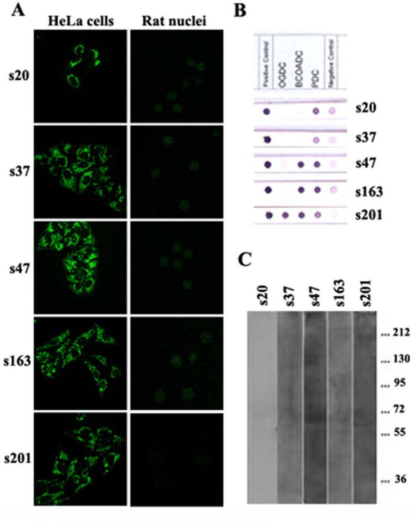
Methods: We screened 103 sera by immunoblotting on nuclear envelopes and indirect immunofluorescence (IIF) using cells and purified nuclei. Reactivities against specific autoantigens were assessed using purified proteins, ELISA, immunoprecipitation and mass spectrometry.
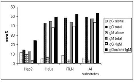
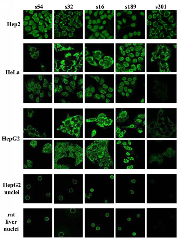
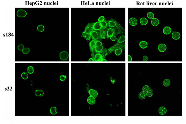
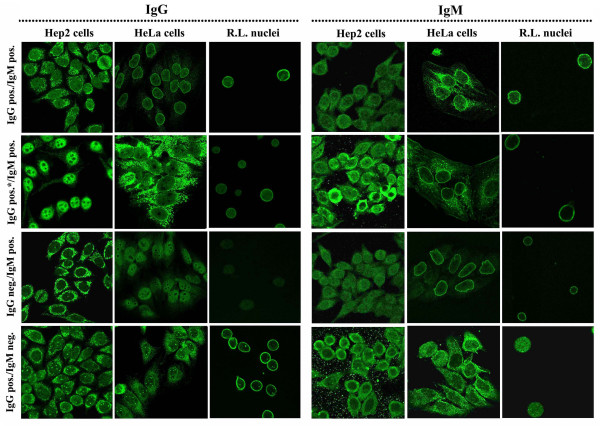
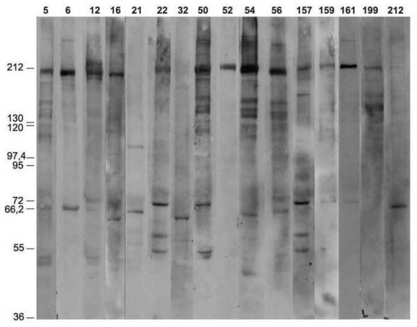
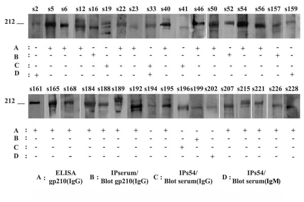
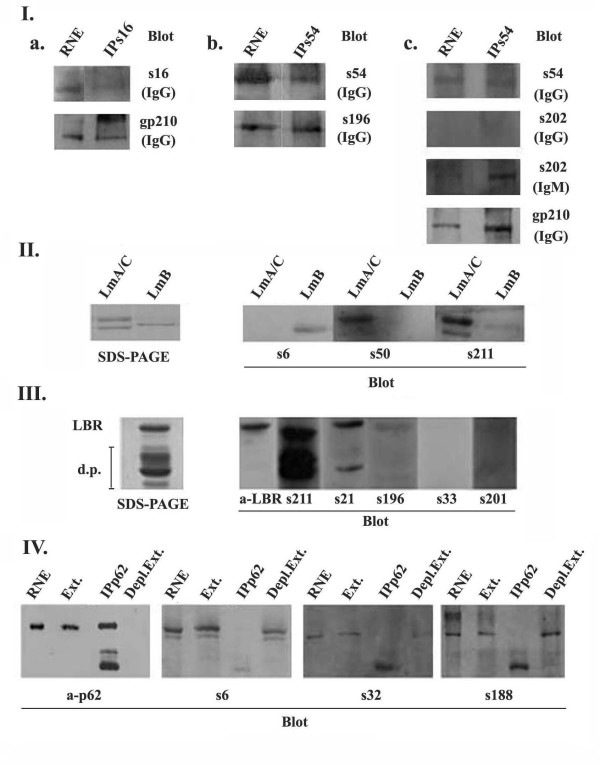
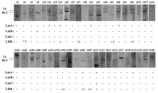
Results: We found higher prevalence of ANEA when sera were assayed by IIF on purified nuclei or cultured cells (50%) compared to Hep2 commercially available slides (15%). Anti-gp210 antibodies were identified in 22.3% and 33% of sera using ELISA for the C-terminal of gp210 or both ELISA and immunoprecipitation, respectively. Immunoblotting on nuclear envelopes revealed that immunoreactivity for the 210 kDa zone is related to anti-gp210 antibodies (p < 0.0001). Moreover, we found that sera had antibodies for lamins A (6.8%), B (1%) and C (1%) and LBR (8.7%), whereas none at all had detectable anti-p62 antibodies.
Conclusions: The prevalence of ANEA or anti-gp210 antibodies is under-estimated in PBC sera which are analyzed by conventional commercially available IIF or ELISA, respectively. Therefore, new substrates for IIF and ELISA should be included by clinical laboratories in the analysis of ANEA in autoimmune sera.
Figures
Similar articles
-
Optimized detection of circulating anti-nuclear envelope autoantibodies by immunofluorescence.BMC Immunol. 2006 Sep 6;7:20. doi: 10.1186/1471-2172-7-20. BMC Immunol. 2006. PMID: 16956395 Free PMC article.
-
Specificity and sensitivity of gp210 autoantibodies detected using an enzyme-linked immunosorbent assay and a synthetic polypeptide in the diagnosis of primary biliary cirrhosis.Hepatology. 1996 May;23(5):1020-4. doi: 10.1002/hep.510230512. Hepatology. 1996. PMID: 8621127 Clinical Trial.
-
Characterization of autoantibodies against components of the nuclear pore complexes: high frequency of anti-p62 nucleoporin antibodies.Ann N Y Acad Sci. 2007 Aug;1109:519-30. doi: 10.1196/annals.1398.058. Ann N Y Acad Sci. 2007. PMID: 17785341
-
Nuclear envelope protein autoantibodies in primary biliary cirrhosis.Semin Liver Dis. 1997 Feb;17(1):79-90. doi: 10.1055/s-2007-1007185. Semin Liver Dis. 1997. PMID: 9089913 Review.
-
Antinuclear antibodies in primary biliary cirrhosis.Semin Liver Dis. 2005 Aug;25(3):298-310. doi: 10.1055/s-2005-916321. Semin Liver Dis. 2005. PMID: 16143945 Review.
Cited by
-
Detection of Autoantibodies Against Nucleoporin p62 in Sera of Patients With Primary Biliary Cholangitis.Ann Lab Med. 2019 May;39(3):291-298. doi: 10.3343/alm.2019.39.3.291. Ann Lab Med. 2019. PMID: 30623621 Free PMC article.
-
Identification of new autoantigens for primary biliary cirrhosis using human proteome microarrays.Mol Cell Proteomics. 2012 Sep;11(9):669-80. doi: 10.1074/mcp.M111.015529. Epub 2012 May 30. Mol Cell Proteomics. 2012. PMID: 22647870 Free PMC article.
-
Diagnostic and Clinical Value of Specific Autoantibodies against Kelch-like 12 Peptide and Nuclear Envelope Proteins in Patients with Primary Biliary Cholangitis.Biomedicines. 2022 Mar 29;10(4):801. doi: 10.3390/biomedicines10040801. Biomedicines. 2022. PMID: 35453551 Free PMC article.
-
Presence of disease specific autoantibodies against liver sinusoidal cells in primary biliary cirrhosis.World J Hepatol. 2013 Oct 27;5(10):568-76. doi: 10.4254/wjh.v5.i10.568. World J Hepatol. 2013. PMID: 24179616 Free PMC article.
-
Expression profile of HMBOX1, a novel transcription factor, in human cancers using highly specific monoclonal antibodies.Exp Ther Med. 2011 May;2(3):487-490. doi: 10.3892/etm.2011.240. Epub 2011 Mar 21. Exp Ther Med. 2011. PMID: 22977529 Free PMC article.
References
-
- Georgatos SD, Theodoropoulos PA. Rules to remodel by: what drives nuclear envelope disassembly and reassembly during mitosis? Crit Rev Eukaryot Gene Expr. 1999;9:373–81. - PubMed
Publication types
MeSH terms
Substances
LinkOut - more resources
Full Text Sources
