Comparative Study
. 2010 May;38(5):403-16.
doi: 10.1016/j.exphem.2010.02.011. Epub 2010 Mar 3.

Runx1 isoforms show differential expression patterns during hematopoietic development but have similar functional effects in adult hematopoietic stem cells

Affiliations
Comparative Study

Runx1 isoforms show differential expression patterns during hematopoietic development but have similar functional effects in adult hematopoietic stem cells

Grant A Challen et al. Exp Hematol. 2010 May.

Abstract

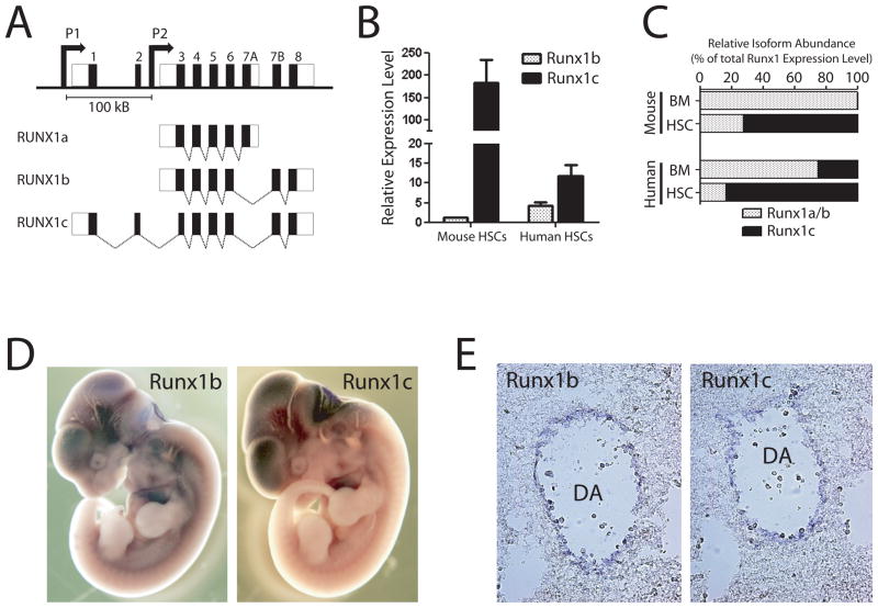
Objective: RUNX1 (also known as acute myeloid leukemia 1) is an essential regulator of hematopoiesis and has multiple isoforms arising from differential splicing and utilization of two promoters. We hypothesized that the rare Runx1c isoform has a distinct role in hematopoietic stem cells (HSCs).

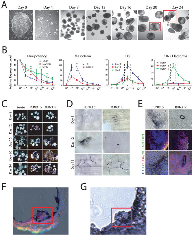
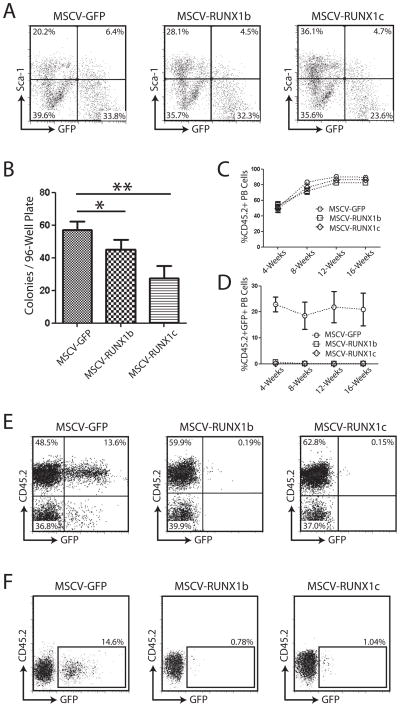
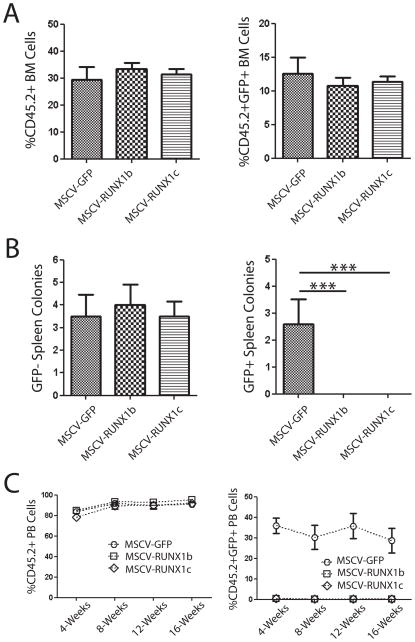
Materials and methods: We have characterized the expression pattern of Runx1c in mouse embryos and human embryonic stem cell (hESC)-derived embryoid bodies using in situ hybridization and expression levels in mouse and human HSCs by real-time polymerase chain reaction. We then determined the functional effects of Runx1c using enforced retroviral overexpression in mouse HSCs.

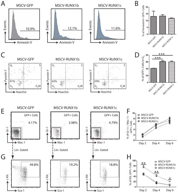
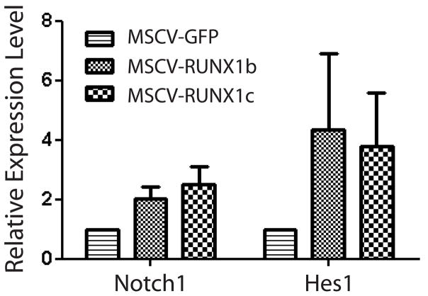
Results: We observed differential expression profiles of RUNX1 isoforms during hematopoietic differentiation of hESCs. The RUNX1a and RUNX1b isoforms were expressed consistently throughout hematopoietic differentiation, whereas the RUNX1c isoform was only expressed at the time of emergence of definitive HSCs. RUNX1c was also expressed in the AGM region of E10.5 to E11.5 mouse embryos, the region where definitive HSCs arise. These observations suggested that the RUNX1c isoform may be important for the specification or function of definitive HSCs. However, using retroviral overexpression to study the effect of RUNX1 isoforms on HSCs in a gain-of-function system, no discernable functional difference could be identified between RUNX1 isoforms in mouse HSCs. Overexpression of both RUNX1b and RUNX1c induced quiescence in mouse HSCs in vitro and in vivo.

Conclusions: Although the divergent expression profiles of Runx1 isoforms during development suggest specific roles for these proteins at different stages of HSC maturation, we could not detect an important functional distinction in adult mouse HSCs using our assay systems.


Conflict of interest statement

Conflict of interest disclosure

The authors have no financial conflicts of interest to disclose.

Figures

Figure 1
Figure 1
RUNX1 genomic locus and isoform expression patterns. (A) Genomic organization of human RUNX1 locus. The RUNX1c isoform is transcribed from the distal P1 promoter and contains the unique N-terminal amino acids encoded by exons 1 and 2. The RUNX1a and predominant RUNX1b isoforms are transcribed from the proximal P2 promoter. The promoters are separated by over 100 Kb in the genome. (B) Expression of RUNX1 isoforms in hematopoietic stem cells. Real-time PCR analysis showed that the RUNX1c isoform is much more highly expressed in HSCs compared to RUNX1b relative to whole bone marrow in both human and mouse. (C) Relative abundance of RUNX1c versus RUNX1a and RUNX1b isoforms in mouse and human bone marrow (BM) and HSCs calculated as a proportional ratio of total RUNX1 expression level. These data show that the RUNX1c isoform is much more abundant in HSCs compared to whole BM. (D) Wholemount in situ hybridization analysis of Runx1 isoform expression patterns in E11.5 mouse embryos. On a gross level, the expression patterns of Runx1 isoforms at this stage of mouse development were highly overlapping. (E) Sectioning of stained embryos showed that Runx1b and Runx1c were both expressed in the endothelium lining the dorsal aorta in the AGM region where definitive HSCs are born.
Figure 2
Figure 2
Modeling human hematopoiesis in vitro with human embryonic stem cell culture. (A) Hematopoietic development in embryoid bodies. Bright field images showing development of hEBs from undifferentiated hESCs (day 0) over time at 4-day intervals. Red squares outline merged inset images. (B) Real-time PCR analysis of hEB differentiation. The pluripotency markers OCT4, NANOG and SOX2 decrease in expression over time as pluripotent hESCs differentiate. The mesoderm markers T and MIXL1 show a dramatic spike in expression between days 4–8 before downregulating, implying that mesoderm is generated between these timepoints in this culture system. The human HSC markers CD34, CD43 and TAL1 peak in expression at day 12 (enclosed by dashed lines) of culture indicating definitive HSCs are being generated during this time window. Analysis of the expression of RUNX1 isoforms during hematopoietic development show that total RUNX1 expression and the RUNX1b isoform are consistent throughout the differentiation process but expression of RUNX1c is dynamic, peaking at day 12 in a similar pattern to that of HSC markers. (C) Wholemount in situ hybridization analysis of RUNX1 isoforms in hEBs. Sense controls show no background staining, but on a gross level no discernable difference can be discriminated between RUNX1b and RUNX1c. (D) Section analysis of stained embryoid bodies showed that at day 12 when there is the most differential expression, RUNX1b is broadly expressed at low levels while RUNX1c is specifically expressed in cells lining the developing cavities in the hEBs. By day 16, the expression profiles of RUNX1b and RUNX1c become highly overlapping. (E) Co-immunofluorescence analysis of stained day 12 hEBs for vascular (VE-CADHERIN) and HSC (CD34) markers. While RUNX1b was broadly expressed throughout the day 12 hEBs, and its expression did not generally correlate with VE-CADHERIN or CD34 expression. RUNX1c however was expressed in a subpopulation of CD34+ cells. (F,G) hEB differentiation in vitro mimics mouse hematopoietic development in vivo. (F) Merged RUNX1b in situ hybridization (purple staining) and immunofluorescence (CD34 – red; VE-CADHERIN – green; DAPI – blue) images of day 20 hEB showing clusters of hematopoietics cells budding off into the lumen. (G) Section of RUNX1b-stained in situ hybridization of AGM region of E11.5 mouse embryo showing presumptive HSCs budding off into the dorsal aorta.
Figure 3
Figure 3
Over-expression of RUNX1 isoforms in mouse hematopoietic progenitor cells. (A) Purification of transduced HSPCs following two days of in vitro culture. All viruses had comparable levels of transduction at this stage. (B) Clonal colony-forming assay for hematopoietic progenitors transduced with RUNX1 isoform retroviruses. Following transduction and culture in vitro for 48-hours, single Sca-1+GFP+ cells were sorted into individual wells of 96-well plates containing Methocult medium. Over-expression of RUNX1 isoforms caused a significant decrease in the number of colonies formed in comparison to cells transduced with MSCV-GFP control virus. (C–E) Peripheral blood analysis of mice transplanted with HSPCs transduced with control (MSCV-GFP) or RUNX1 isoform retroviruses. There was no difference in the total number of donor-derived (CD45.2+) peripheral blood cells between the three transplant groups (C), but there was a marked difference in the number of GFP+ cells generated from transduced cells (D) with virtually no generation of progeny from stem/progenitor cells over-expressing RUNX1 isoforms. (E) Representative examples of peripheral blood analysis of mice transplanted with HSPCs transduced with retroviruses 4-weeks post-transplant. While robust GFP expression was detected in peripheral blood cells of control mice (MSCV-GFP), virtually no GFP+ cells were detected in mice transplanted with cells over-expressing RUNX1 isoforms. (F) Analysis of the HSC compartment of long-term transplanted mice. Analysis of the SPKLS fraction from the bone marrow of mice transplanted with hematopoietic progenitors transduced with retrovirus showed that some GFP+ RUNX1 over-expressing HSCs could be detected >16-weeks post-transplant. Although the percentage was greatly reduced compared to control animals, this suggests that over-expression of RUNX1 isoforms in mouse HSCs was not toxic but induced extreme quiescence.
Figure 4
Figure 4
(A) Over-expression of RUNX1 isoforms in mouse hematopoietic stem/progenitor cells does not affect homing to the bone marrow. Analysis of the bone marrow of recipient mice 24-hours post-transplant of transduced hematopoietic progenitors showed that there was no difference in the total number of donor cells (CD45.2+) or transduced donor cells (CD45.2+GFP+) in the bone marrow of recipient mice. (B) The spleen CFU-S colony assay demonstrated that although there was no difference in the number of GFP spleen colonies in mice transplanted with transduced cells, no GFP+ colonies were detected from cells over-expressing RUNX1 isoforms. (C) Over-expression of RUNX1 isoforms in fetal liver HSCs. Transplantation of transduced fetal liver HSCs showed that while there was no difference in total number of donor-derived peripheral blood cells (CD45.2+), essentially no peripheral blood progeny (CD45.2+GFP+) could be detected from HSCs over-expressing RUNX1 isoforms.
Figure 5
Figure 5
(A) Examples of AnnexinV staining for apoptosis in retrovirally transduced hematopoietic stem/progenitor cells. (B) No difference was detected in the number of RUNX1 over-expressing cells undergoing apoptosis compared to control cells (MSCV-GFP). (C) Examples of Hoechst/Pyronin Y staining of retrovirally transduced HSPCs following two days of in vitro culture. (D) Over-expression of RUNX1 isoforms in HSPCs caused an inhibition of proliferation in vitro with a significantly higher proportion of cells in G0 or quiescence. (E) Examples of flow cytometric analysis of transduced HSPCs after four days culture for the differentiation markers Mac-1 and Gr-1. (F) Over-expression of RUNX1-isoforms in HSPCs did not increase the rate of differentiation compared to MSCV-GFP transduced control. (G) Examples of flow cytometric analysis of transduced HSPCs after four days culture for the hematopoietic progenitor compartment (KSL). (H) Over-expression of RUNX1-isoforms resulted in a significant reduction in the primitive progenitor compartment from transduced cells.
Figure 6
Figure 6
Real-time PCR analysis of retrovirally transduced hematopoietic stem/progenitor cells for Notch pathway genes. Over-expression of RUNX1 isoforms induced an upregulation of the surface receptor Notch1 and the downstream target gene Hes1 compared to control cells (MSCV-GFP).

Similar articles

Cited by

References

    1. Look AT. Oncogenic transcription factors in the human acute leukemias. Science. 1997;278:1059–1064. - PubMed
    1. Speck NA, Gilliland DG. Core-binding factors in haematopoiesis and leukaemia. Nat Rev Cancer. 2002;2:502–513. - PubMed
    1. Okuda T, van Deursen J, Hiebert SW, Grosveld G, Downing JR. AML1, the target of multiple chromosomal translocations in human leukemia, is essential for normal fetal liver hematopoiesis. Cell. 1996;84:321–330. - PubMed
    1. Wang Q, Stacy T, Binder M, Marin-Padilla M, Sharpe AH, Speck NA. Disruption of the Cbfa2 gene causes necrosis and hemorrhaging in the central nervous system and blocks definitive hematopoiesis. Proc Natl Acad Sci U S A. 1996;93:3444–3449. - PMC - PubMed
    1. Okada H, Watanabe T, Niki M, et al. AML1(−/−) embryos do not express certain hematopoiesis-related gene transcripts including those of the PU.1 gene. Oncogene. 1998;17:2287–2293. - PubMed

Publication types

MeSH terms