. 2010 May 19;167(3):644-55.
doi: 10.1016/j.neuroscience.2010.02.048. Epub 2010 Mar 3.

Developmental regulation and neuroprotective effects of striatal tonic GABAA currents

Affiliations

Developmental regulation and neuroprotective effects of striatal tonic GABAA currents

V Santhakumar et al. Neuroscience. .

Abstract

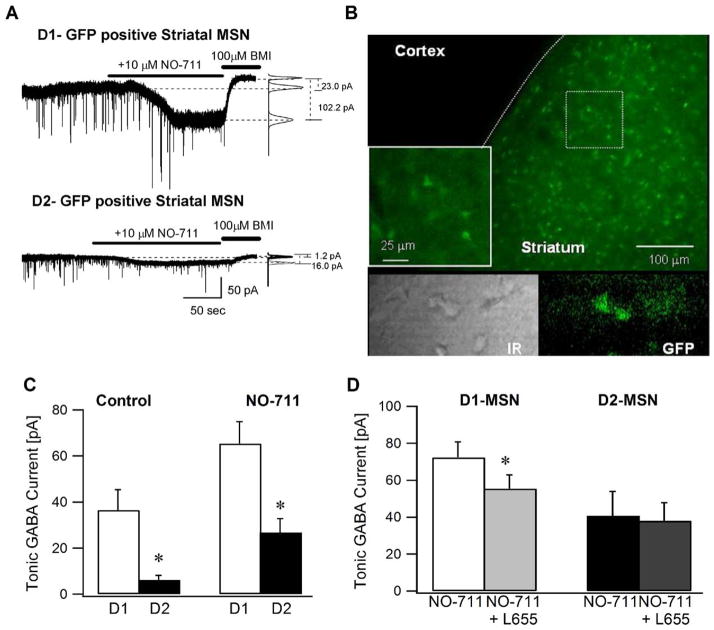
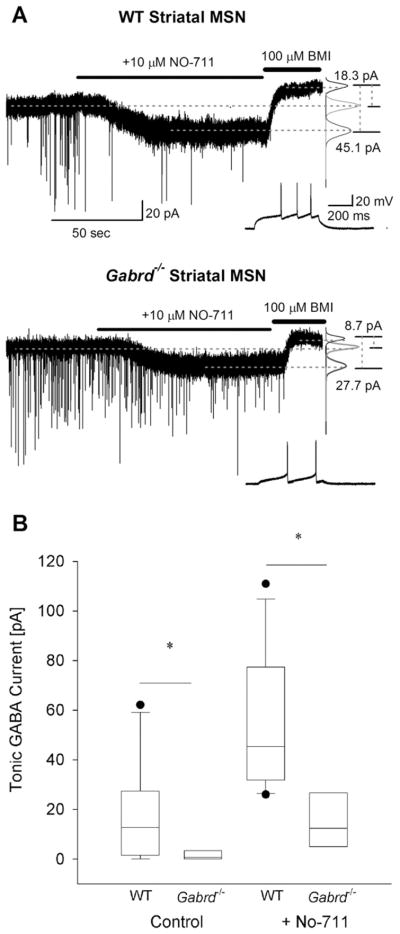
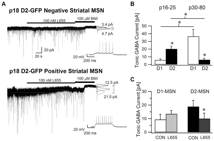
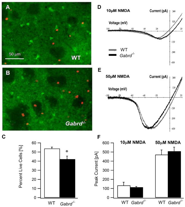
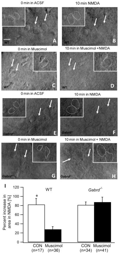
Striatal neurons are known to express GABA(A) receptor subunits that underlie both phasic and tonic inhibition. Striatal projection neurons, or medium spiny neurons (MSNs), are divided into two classes: MSNs containing the dopamine D1 receptor (D1-MSNs) form the direct pathway to the substantia nigra and facilitate movement while MSNs expressing the dopamine D2 receptor (D2-MSNs) form the pallidal pathway that inhibits movement. Consequently, modulating inhibition in distinct classes of MSNs will differentially impact downstream network activity and motor behavior. Given the powerful role of extrasynaptic inhibition in controlling neuronal excitability, we examined the nature of striatal tonic inhibition and its potential role in preventing excitotoxicity. Consistent with earlier studies in young (P16-P25) mice, tonic GABA currents in D2-MSNs were larger than in D1-MSNs. However, with age (>P30 mice) the tonic GABA currents increased in D1-MSNs but decreased in D2-MSNs. These data demonstrate a developmental switch in the MSN subtype expressing larger tonic GABA currents. Compared to wild-type, MSNs from adult mice lacking the GABA(A)R delta subunit (Gabrd(-/-) mice) had both decreased tonic GABA currents and reduced survival following an in vitro excitotoxic challenge with quinolinic acid. Furthermore, muscimol-induced tonic GABA currents were accompanied by reduced acute swelling of striatal neurons after exposure to NMDA in WT mice but not in Gabrd(-/-) mice. Our data are consistent with a role for tonic inhibition mediated by GABA(A)R delta subunits in neuroprotection against excitotoxic insults in the adult striatum.


Figures

Fig. 1
Fig. 1
Adult striatal medium spiny neurons expressing D1 receptors have larger tonic GABA currents. (A) Representative voltage-clamp recordings (Vh=− 70 mV) from adult D1-MSNs (above) and D2-MSNs (below) illustrate the magnitude of tonic GABA current blocked by a saturating concentration of BMI (100 μM). Panels to the right show Gaussian fits to all-points histograms derived from 30 s recording periods in control conditions and in the presence of the GABA transporter-1 blocker NO-711 (10 μM) and a 15 s recording period during the perfusion of BMI used to determine the tonic current. The dashed lines indicate the Gaussian means and the difference currents are noted. (B) Confocal image of a cortico–striatal slice from a D2-GFP mouse shows that the GFP expression is restricted to the striatum. Inset illustrates a magnified image of the boxed area. The lower panel shows an IR-DIC (left) and fluorescence image (right) of a representative cortico-striatal slice from a D2-GFP mouse. (C) Summary histogram of the tonic GABA currents in D1- and D2-MSNs under control conditions and in the presence of the GABA transporter antagonist NO-711. (D) Histogram shows the effect of L655,708 (100 nM), an inverse benzodiazepine site agonist selective for GABAAR containing the α5 subunit on tonic inhibition in adult D1- and D2-MSNs in the presence of 10 μM NO-711. Tonic GABA currents were recorded in the presence of glutamate receptor blockers and 5 μM GABA. Asterisk denotes a statistically significant (P<0.05) difference in mean values using Student t-test.
Fig. 2
Fig. 2
Tonic GABA currents are decreased in Gabrd−/− mice lacking GABAA receptor delta subunits. (A) Example voltage-clamp traces from striatal MSNs in adult wild-type (above) and Gabrd−/− mice (below) show the difference in tonic GABA currents in control conditions and in the presence of NO-711 (10 μM) between the two genotypes. Recordings were obtained at a holding potential of −70 mV. To the right are panels showing Gaussian fits to all-points histograms derived from 30 s recording periods in control conditions and in the presence of NO-711 (10 μM) and a 15 s recording period during the perfusion of BMI used to determine the tonic current. The difference currents are noted to the right were calculated from the Gaussian means indicated by the dashed lines. Inset shows the characteristic firing pattern of the MSNs recorded in response to a depolarizing current injection (+80 pA in the WT in the upper panel and +20 pA in Gabrd−/− in the lower panel) from a holding potential of −70 mV. (B) Summary box-plot shows the decrease in tonic GABA currents in MSNs from Gabrd−/− mice in the absence and presence of NO-711. The upper and lower sides of the box represent the upper/lower quartiles and the median value is represented as a line. The maximum and minimum values are represented by the extent of the whiskers with outliers shown as black dots. Asterisk denotes a statistically significant (P<0.05) difference in mean values using Student’s t-test. Recordings were obtained in the presence of glutamate receptor blockers and 5 μM GABA.
Fig. 3
Fig. 3
Striatal medium spiny neurons expressing D2 receptors have larger tonic GABA currents in young mice. (A) Recordings from a representative D1-MSN (above) and D2-MSN (below) from juvenile (P18) D2-GFP mice show the tonic GABA current blocked by a saturating concentration of BMI (100 μM). The effect of the α5 selective GABAA receptor inverse agonist, L655,708 (100 nM) on tonic GABA currents in D1 and D2-MSN is also illustrated. As before, the graphs to the right show Gaussian fits to all-points histograms derived from 30 s periods in control and L655,708 and a 15 s recording in BMI with dashed lines indicating the means used to calculate the tonic current. Inset shows the characteristic firing pattern of the MSN recorded in response to a depolarizing current injection (+120 pA) from a holding potential of −70 mV. (B) Summary data compare the tonic GABA currents in juvenile D1- and D2-MSNs with the tonic GABA currents from adult D1 and D2-MSNs (same as control data as in Fig. 1C) under control conditions in the presence of glutamate receptor blockers and 5 μM GABA. (C) Histogram shows the effect of L655,708 (100 nM) on tonic GABA currents in juvenile D1- and D2-MSNs. All recordings were performed at a holding potential at −70 mV. Asterisk denotes a statistically significant (P<0.05) difference in mean values.
Fig. 4
Fig. 4
Enhanced susceptibility of striatal MSNs in Gabrd−/− mice to excitotoxic cell death. (A, B) Confocal photomicrograph of cortico–striatal slices from a wild-type mouse (A) and a Gabrd−/− mouse (B) showing the assay for live and dead cells following in vitro exposure to quinolinic acid (0.5 mM, 1 h). The live cells are stained with Calcein (in green) and dead cells incorporate the nuclear stain Ethidium Homodimer-1 (in red). Both Calcein and Ethidium Homodimer-1 images of a given field of view were obtained at 10× using appropriate filters and overlaid for illustration. (C) Summary data show the percent of MSNs that survive an hour long in vitro exposure to the excitotoxin quinolinic acid (0.5 mM) in wild-type and Gabrd−/− mice. (D, E) Representative NMDA current–voltage plots obtained in MSNs from adult wild-type and Gabrd−/− mice illustrate comparable peak NMDA currents and voltage-dependence in the presence 10 μM (D) and 50 μM (E) NMDA. (F) Summary histogram shows that the peak NMDA currents are similar in MSNs of wild type and Gabrd−/− mice. Asterisk denote a statistically significant (P<0.05) difference.
Fig. 5
Fig. 5
Enhancement of tonic currents mediated by δ subunit-containing GABAARs decreases excitotoxic cell swelling of MSNs. (A–D). Illustrative IR-DIC images of wild-type striatal MSN obtained using a 40× objective show the MSNs before (A) and 10 min after exposure 50 μM NMDA (B) show the swelling and indistinct outlines after NMDA exposure in aCSF (n =17 cells from six slices in three mice). Images from slices in which tonic inhibition was selectively enhanced by a 5 min perfusion of 50 nM muscimol before (C) and during the 10 min exposure to 50 μM NMDA (D) show that muscimol decreases NMDA induced swelling in MSNs (n =36 cells from 10 slices in four mice). Insets show outlines used to measure the area and perimeter of the cells indicated by arrows. (E–H). Images of striatal MSNs from Gabrd−/− mice before (E) and 10 min after exposure 50 μM NMDA (F) show the NMDA induced swelling (n =34 cells from nine slices in three mice). Images obtained following enhancement of tonic inhibition by muscimol before (G) and during the 10 min exposure to NMDA (H) show that muscimol does not protect against NMDA induced swelling of MSNs in Gabrd−/− mice (n =41 cells from 10 slices in three mice). Insets show outlines used to measure the area and perimeter of the cells indicated by the arrows. Scale bar in (A) represents 10 μm. (I) Summary histogram shows the degree of NMDA induced increase in cell area in wild-type and Gabrd−/− mice in the absence and presence of muscimol. Asterisk denotes a statistically significant (P<0.05) difference.

Similar articles

Cited by

References

    1. Ade KK, Janssen MJ, Ortinski PI, Vicini S. Differential tonic GABA conductances in striatal medium spiny neurons. J Neurosci. 2008;28:1185–1197. - PMC - PubMed
    1. Albin RL, Reiner A, Anderson KD, Dure LS, Handelin B, Balfour R, Whetsell WO, Jr, Penney JB, Young AB. Preferential loss of striato-external pallidal projection neurons in presymptomatic Huntington’s disease. Ann Neurol. 1992;31:425–430. - PubMed
    1. Balleine BW, Delgado MR, Hikosaka O. The role of the dorsal striatum in reward and decision-making. J Neurosci. 2007;27:8161–8165. - PMC - PubMed
    1. Beister A, Kraus P, Kuhn W, Dose M, Weindl A, Gerlach M. The N-methyl-D-aspartate antagonist memantine retards progression of Huntington’s disease. J Neural Transm Suppl. 2004;68:117–122. - PubMed
    1. Bracci E, Panzeri S. Excitatory GABAergic effects in striatal projection neurons. J Neurophysiol. 2006;95:1285–1290. - PubMed

Publication types

MeSH terms