

MnSOD activity protects mitochondrial morphology of quiescent fibroblasts from age associated abnormalities
- PMID: 20206302
- PMCID: PMC2874624
- DOI: 10.1016/j.mito.2010.02.004
MnSOD activity protects mitochondrial morphology of quiescent fibroblasts from age associated abnormalities
Abstract
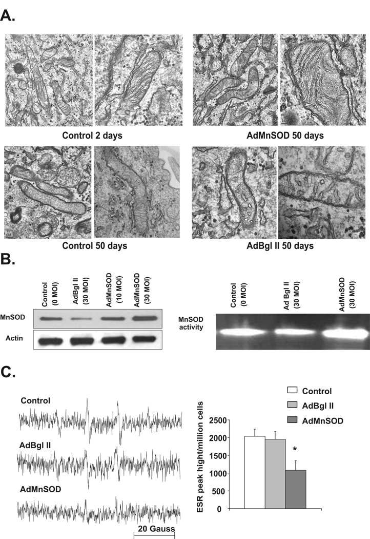
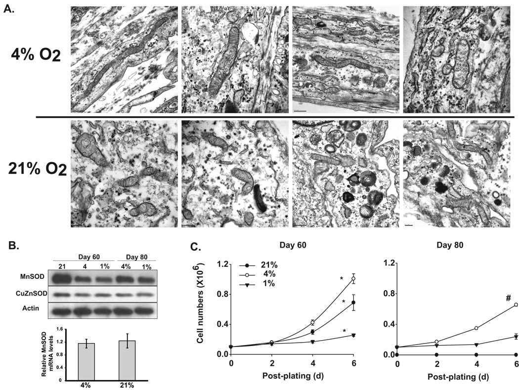
Previously, we have shown manganese superoxide dismutase (MnSOD) activity protects quiescent human normal skin fibroblasts (NHFs) from age associated loss in proliferative capacity. The loss in proliferative capacity of aged vs. young quiescent cells is often characterized as the chronological life span, which is clearly distinct from replicative senescence. We investigate the hypothesis that MnSOD activity protects the mitochondrial morphology from age associated damage and preserves the chronological life span of quiescent fibroblasts. Aged quiescent NHFs exhibited abnormalities in mitochondrial morphology including abnormal cristae formation and increased number of vacuoles. These results correlate with the levels of cellular reactive oxygen species (ROS) and mitochondrial morphology in MnSOD homozygous and heterozygous knockout mouse embryonic fibroblasts. The abnormalities in mitochondrial morphology in aged quiescent NHFs cultured in presence of 21% oxygen concentration were more severe than NHFs cultured in 4% oxygen environment. The alteration in mitochondrial morphology was associated with a significant increase in cell population doubling: 54h in 21% compared to 44h in 4% oxygen environment. Overexpression of MnSOD decreased ROS levels, and preserved mitochondrial morphology in aged quiescent NHFs. These results demonstrate that MnSOD activity protects mitochondrial morphology and preserves the proliferative capacities of quiescent NHFs from age associated loss.
Copyright 2010 Mitochondria Research Society. Published by Elsevier B.V. All rights reserved.
Figures

Similar articles
-
MnSOD activity regulates hydroxytyrosol-induced extension of chronological lifespan.Age (Dordr). 2012 Feb;34(1):95-109. doi: 10.1007/s11357-011-9223-7. Epub 2011 Mar 8. Age (Dordr). 2012. PMID: 21384152 Free PMC article.
-
N-acetyl-L-cysteine increases MnSOD activity and enhances the recruitment of quiescent human fibroblasts to the proliferation cycle during wound healing.Mol Biol Rep. 2016 Jan;43(1):31-9. doi: 10.1007/s11033-015-3935-1. Epub 2015 Dec 15. Mol Biol Rep. 2016. PMID: 26671656 Free PMC article.
-
Manganese superoxide dismutase activity regulates transitions between quiescent and proliferative growth.Aging Cell. 2008 Jun;7(3):405-17. doi: 10.1111/j.1474-9726.2008.00384.x. Epub 2008 Mar 10. Aging Cell. 2008. PMID: 18331617 Free PMC article.
-
Manganese superoxide dismutase regulates a redox cycle within the cell cycle.Antioxid Redox Signal. 2014 Apr 1;20(10):1618-27. doi: 10.1089/ars.2013.5303. Epub 2013 May 29. Antioxid Redox Signal. 2014. PMID: 23590434 Free PMC article. Review.
-
Managing odds in stem cells: insights into the role of mitochondrial antioxidant enzyme MnSOD.Free Radic Res. 2016;50(5):570-84. doi: 10.3109/10715762.2016.1155708. Free Radic Res. 2016. PMID: 26899340 Review.
Cited by
-
Molecular characterization and expression analysis of iron superoxide dismutase gene from Pseudochlorella pringsheimii (Trebouxiophyceae, Chlorophyta).Physiol Mol Biol Plants. 2019 Jan;25(1):221-228. doi: 10.1007/s12298-018-0569-5. Epub 2018 Jun 18. Physiol Mol Biol Plants. 2019. PMID: 30804644 Free PMC article.
-
Arachidonate 12-lipoxygenase and 12-hydroxyeicosatetraenoic acid contribute to stromal aging-induced progression of pancreatic cancer.J Biol Chem. 2020 May 15;295(20):6946-6957. doi: 10.1074/jbc.RA120.012798. Epub 2020 Apr 7. J Biol Chem. 2020. PMID: 32265301 Free PMC article.
-
MnSOD activity regulates hydroxytyrosol-induced extension of chronological lifespan.Age (Dordr). 2012 Feb;34(1):95-109. doi: 10.1007/s11357-011-9223-7. Epub 2011 Mar 8. Age (Dordr). 2012. PMID: 21384152 Free PMC article.
-
Involvement of oxidative stress and mitochondrial mechanisms in air pollution-related neurobiological impairments.Neurobiol Stress. 2019 Dec 19;12:100205. doi: 10.1016/j.ynstr.2019.100205. eCollection 2020 May. Neurobiol Stress. 2019. PMID: 32258254 Free PMC article.
-
Mitofusin 1 and optic atrophy 1 shift metabolism to mitochondrial respiration during aging.Aging Cell. 2017 Oct;16(5):1136-1145. doi: 10.1111/acel.12649. Epub 2017 Jul 31. Aging Cell. 2017. PMID: 28758339 Free PMC article.
References
-
- Asikainen TM, Huang TT, et al. Increased sensitivity of homozygous Sod2 mutant mice to oxygen toxicity. Free Radic. Biol. Med. 2002;32:175–186. - PubMed
-
- Balaban RS, Nemoto S, Finkel T. Mitochondria, oxidants, and aging. Cell. 2005;120:483–495. - PubMed
-
- Beauchamp C, Fridovich I. Superoxide dismutase: improved assays and an assay applicable to acrylamide gels. Anal. Biochem. 1971;44:276–287. - PubMed
-
- Borgstahl GE, Parge HE, et al. The structure of human mitochondrial manganese superoxide dismutase reveals a novel tetrameric interface of two 4-helix bundles.[erratum appears in Cell 1993 Feb 12;72(3):following 476] Cell. 1992;71:107–118. - PubMed
Publication types
MeSH terms
Substances
Grants and funding
LinkOut - more resources
Full Text Sources
