. 2010 Apr;52(4):586-93.
doi: 10.1016/j.jhep.2010.01.003. Epub 2010 Feb 13.

Palmitoleate attenuates palmitate-induced Bim and PUMA up-regulation and hepatocyte lipoapoptosis

Affiliations

Palmitoleate attenuates palmitate-induced Bim and PUMA up-regulation and hepatocyte lipoapoptosis

Yuko Akazawa et al. J Hepatol. 2010 Apr.

Abstract

Background & aims: Saturated free fatty acids induce hepatocyte lipoapoptosis. This lipotoxicity involves an endoplasmic reticulum stress response, activation of JNK, and altered expression and function of Bcl-2 proteins. The mono-unsaturated free fatty acid palmitoleate is an adipose-derived lipokine which suppresses free fatty acid-mediated lipotoxicity by unclear mechanisms. Herein we examined the mechanisms responsible for cytoprotection.

Methods: We employed isolated human and mouse primary hepatocytes, and the Huh-7 and Hep 3B cell lines for these studies. Cells were incubated in presence and absence of palmitate (16:0), stearate (18:0), and or palmitoleate (16:1, n-7).

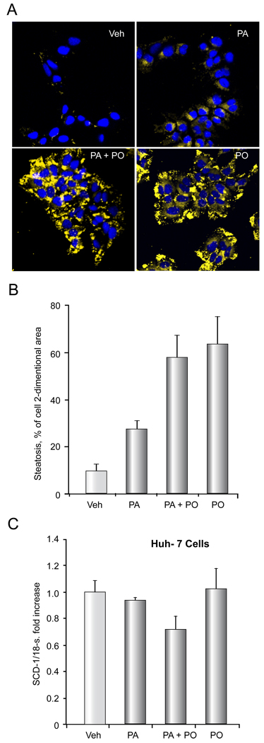
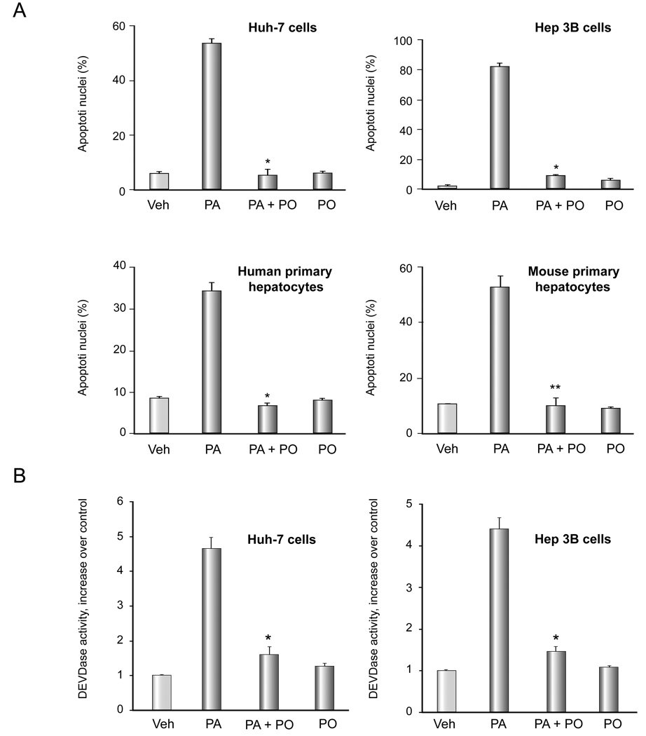
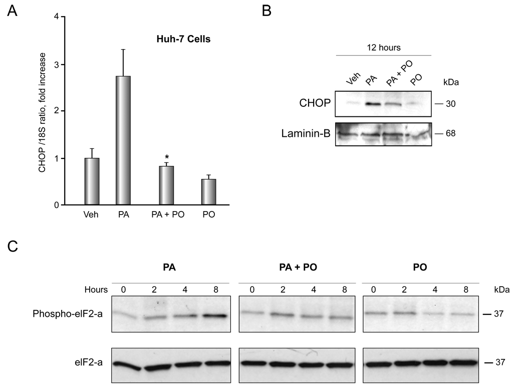
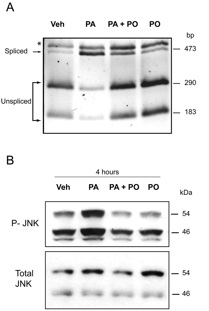
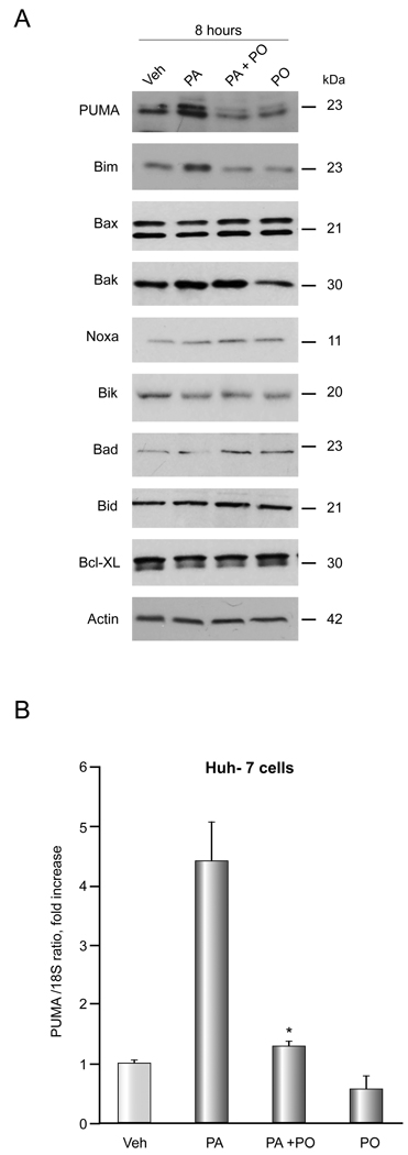
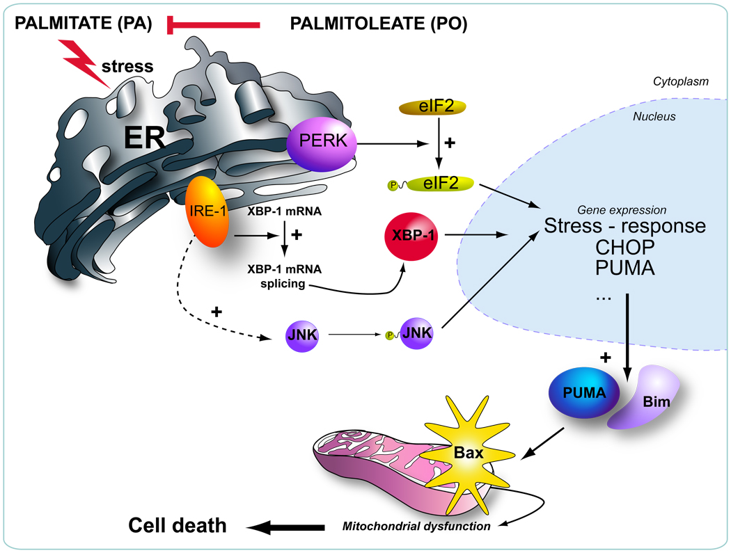
Results: Palmitoleate significantly reduced lipoapoptosis by palmitate or stearate in both primary cells and cell lines. Palmitoleate accentuated palmitate-induced steatosis in Huh-7 cells excluding inhibition of steatosis as a mechanism for reduced apoptosis. Palmitoleate inhibited palmitate induction of the endoplasmic reticulum stress response as demonstrated by reductions in CHOP expression, eIF2-alpha phosphorylation, XBP-1 splicing, and JNK activation. Palmitate increased expression of the BH3-only proteins PUMA and Bim, which was attenuated by palmitoleate. Consistent with its inhibition of PUMA and Bim induction, palmitoleate prevented activation of the downstream death mediator Bax.

Conclusions: These data suggest palmitoleate inhibits lipoapoptosis by blocking endoplasmic reticulum stress-associated increases of the BH3-only proteins Bim and PUMA.


Figures

Figure 1
Figure 1. PO does not attenuate PA- mediated steatosis
Nile red staining was performed on Huh-7 cells treated with vehicle (Veh), 200 µM PA, 200 µM PO plus 200 µM PA, or 200 µM PO alone for 18 h. (A) Representative fluorescent photomicrographs (× 60) are depicted. Nile red fluoresces as a yellow-gold at about 510nm; cells were counter-stained for nucleic acids by DAPI (blue). (B), Cellular steatosis was quantified in 5 random low power fields for each condition with image analysis software. Total area of lipid per cell (pixels above threshold) is represented. The data represent the mean ± SEM for n = 3 studies. (C) Huh-7 cells were treated with vehicle (Veh), 800 µM PA, 400 µM PO plus 800 µM PA, or 400 µM PO alone for 8 hours. SCD-1 mRNA was quantified by real time PCR. Fold induction was determined by normalization to 18S ribosomal RNA. Data represent the mean ± SEM of 3 independent experiments.
Figure 2
Figure 2. PO attenuates PA-mediated apoptosis
(A) Cells were treated with free fatty acids. The concentration of PA was 800 µM for Huh-7 cell, 400uM for Hep 3B cells and 200 µM for primary hepatocytes. The ratio of PA: PO was 2:1 for Huh-7 cells and 1:1 for the remainder of the cell types. Apoptosis was assessed by morphological criteria after DAPI staining. The data represent the mean ± SEM for n = 3 studies. * p < 0.01, ** p < 0.05, PA-treated cells vs. PA plus PO-treated cells. (B) After Huh-7 cells were treated as in (A), activity of effector caspases 3 and 7 was measured by a fluorogenic assay. Data are expressed as fold-increase of relative fluorescence units (RFLU) over control value (untreated cells), which was arbitrarily set to 1, and represent the mean ± SEM for n=3 studies each performed in triplicate. * p<0.05, PA-treated cells vs. PA plus PO-treated cells.
Figure 3
Figure 3. PO inhibits PA-induced CHOP up-regulation and phosphorylation of eIF2-α
(A) Huh-7 cells were treated with vehicle (Veh), 800 µM PA, 800 µM PA plus 400 µM PO, or 400 µM PO PO alone for 8 hours. CHOP mRNA was quantified by real time PCR. Fold induction was determined by normalization to 18S. Data represent the mean and error of 3 independent experiments each performed in triplicate. * p < 0.05, PA-treated cells vs. PA plus PO-treated cells. (B) Huh-7 cells were treated as above. Nuclear extracts were obtained from each experimental conditions and were subjected to immunoblot analysis for CHOP. Lamin B was used as a loading control. (C) Huh-7 cells were treated as above for the indicated time. Whole cell lysates were obtained and resolved by SDS-PAGE. Proteins were identified by western blot analysis by using antisera specific for total and phospho-eIF2-α.
Figure 4
Figure 4. PO inhibits PA-induced XBP-1 and JNK activation
(A) Huh-7 cells were treated with either vehicle (Veh), 800 µM PA, 800 µM PA plus 400 µM PO, or 400 µM PO alone for 8 hours. XBP-1 cDNA was amplified by PCR followed by 1 hourincubation with Pst1. Unspliced form of XBP-1 shows 290-bp and 180-bp product (which are mainly observed in Veh, PA+PO, PO-treated cells). Spliced form shows single 473-bp amplification product (which is mainly observed in PA-treated cells). * Undigested PCR product. (B) Huh-7 cells were treated as above for 4 hours. Whole cell lysates were obtained and resolved by SDS-PAGE. Proteins were identified by western blot analysis by using antisera specific for total and active (phospho) members of JNK 1/2.
Figure 5
Figure 5. PO inhibits PA-mediated Bim and PUMA induction and Bax activation
(A) (A, B) Western blot analysis was performed for Bcl-2 proteins using whole cell lysates from Huh-7 cells treated with vehicle (Veh), 800 µM PA, 800 µM PA plus 400 µM PO, or 400 µM PO alone for 8 hours. (B) Huh-7 cells were treated with FFAs as described above. PUMA mRNA was quantified by real time PCR. Fold induction was determined by normalization to 18S. Data represent the mean and error of 3 independent experiments each performed in triplicate. * p < 0.05, PA-treated cells vs. PA plus PO-treated cells. (C) Huh-7 cells were examined by immunofluorescence microscopy for activation of Bax following treatment with either vehicle (Veh), 800 µM PA, 800 µM PA plus 400 µM PO, or 400 µM PO alone for 16 hours. The primary antibody employed recognizes the N-terminus of Bax which is exposed as an antigen upon Bax activation. Green fluorescence shows activated Bax and blue fluorescence shows DAPI staining. The graph shows quantification of 5 experiments. * p < 0.01, PA-treated cells vs. PA plus PO-treated cells.
Figure 6
Figure 6. Schematic representation of the proposed model for the mechanism of PO inhibiting PA-induced apoptosis
Palmitoleate (PO) inhibits palmitate (PA)-induced ER stress, thereby preventing induction of the BH3-only proteins PUMA, Bim, and Bax activation.

Similar articles

Cited by

References

    1. Angulo P. Nonalcoholic fatty liver disease. N Engl J Med. 2002;346:1221–1231. - PubMed
    1. Parekh S, Anania FA. Abnormal lipid and glucose metabolism in obesity: implications for nonalcoholic fatty liver disease. Gastroenterology. 2007;132:2191–2207. - PubMed
    1. Unger RH, Orci L. Lipoapoptosis: its mechanism and its diseases. Biochim Biophys Acta. 2002;1585:202–212. - PubMed
    1. Malhi H, Gores GJ. Molecular mechanisms of lipotoxicity in nonalcoholic fatty liver disease. Semin Liver Dis. 2008;28:360–369. - PMC - PubMed
    1. Feldstein AE, Gores GJ. Apoptosis in alcoholic and nonalcoholic steatohepatitis. Front Biosci. 2005;10:3093–3099. - PubMed

Publication types

MeSH terms

Substances