. 2010 May 15;341(2):346-59.
doi: 10.1016/j.ydbio.2010.02.024. Epub 2010 Mar 4.

Synovial joint morphogenesis requires the chondrogenic action of Sox5 and Sox6 in growth plate and articular cartilage

Affiliations

Synovial joint morphogenesis requires the chondrogenic action of Sox5 and Sox6 in growth plate and articular cartilage

Peter Dy et al. Dev Biol. .

Abstract

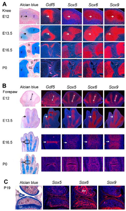
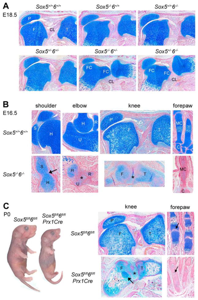
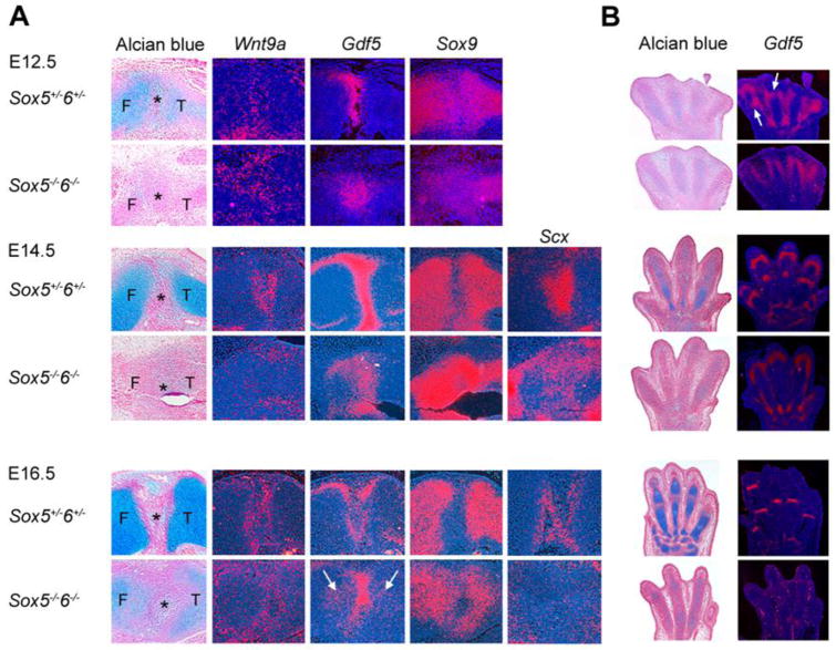
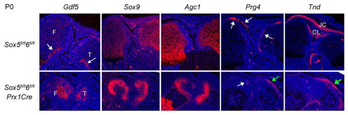
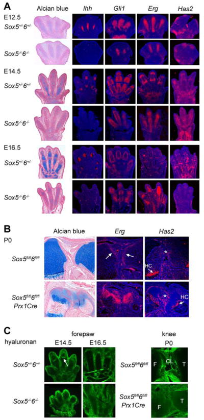
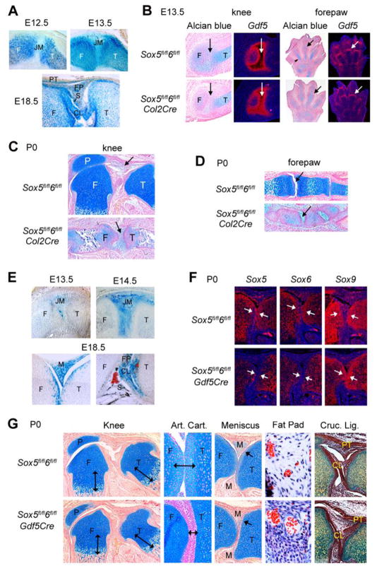
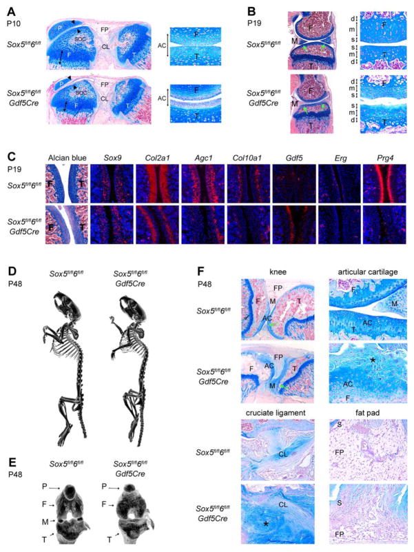
The mechanisms underlying synovial joint development remain poorly understood. Here we use complete and cell-specific gene inactivation to identify the roles of the redundant chondrogenic transcription factors Sox5 and Sox6 in this process. We show that joint development aborts early in complete mutants (Sox5(-/-)6(-/-)). Gdf5 and Wnt9a expression is punctual in articular progenitor cells, but Sox9 downregulation and cell condensation in joint interzones are late. Joint cell differentiation is unsuccessful, regardless of lineage, and cavitation fails. Sox5 and Sox6 restricted expression to chondrocytes in wild-type embryos and continued Erg expression and weak Ihh expression in Sox5(-/-)6(-/-) growth plates suggest that growth plate failure contribute to this Sox5(-/-)6(-/-) joint morphogenesis block. Sox5/6 inactivation in specified joint cells and chondrocytes (Sox5(fl/fl)6(fl/fl)Col2Cre) also results in a joint morphogenesis block, whereas Sox5/6 inactivation in specified joint cells only (Sox5(fl/fl)6(fl/fl)Gdf5Cre) results in milder joint defects and normal growth plates. Sox5(fl/fl)6(fl/fl)Gdf5Cre articular chondrocytes remain undifferentiated, as shown by continued Gdf5 expression and pancartilaginous gene downregulation. Along with Prg4 downregulation, these defects likely account for joint tissue overgrowth and incomplete cavitation in adult mice. Together, these data suggest that synovial joint morphogenesis relies on essential roles for Sox5/6 in promoting both growth plate and articular chondrocyte differentiation.


Figures

Fig. 1
Fig. 1
Expression of Sox5, Sox6, and Sox9 in synovial joints. Adjacent sections through the knee (A), forepaw (B), and meniscus (C) of wild-type mice at various ages were stained with Alcian blue or hybridized with Gdf5 and Sox RNA probes, as indicated. The arrows in (A) point to the presumptive knee joint. The double-headed arrows in (B) mark digital condensations, whereas the arrows point to a developing phalangeal joint. The RNA expression data at E16.5 and P0 are shown as high-magnification pictures of the area boxed in the sections stained with Alcian blue. Note in (C) that Sox6 is strongly expressed in bone marrow (BM). AC, articular cartilage; CL, cruciate ligament; F, femur; FP, fat pad; GP, growth plate. M, meniscus; T, tibia; S, synovium and fat pad; PT, patellar tendon.
Fig. 2
Fig. 2
Analysis of developing synovial joints in mice harboring Sox5 and Sox6 null alleles. (A) Mid-sagittal sections through the knees of E18.5 fetuses harboring various combinations of Sox5 and Sox6 null alleles. Note that although all sections are mid-sagittal, as indicated by the presence of the patella (P), the two femoral condyles (FC) are seen and cruciate ligaments (CL) are stretched in the Sox5/6+/ and Sox5+/6/ sections, reflecting valgus deformation. F, femur; T, tibia. (B) Sections through the shoulder, elbow, knee and forepaw of E16.5 wild-type and Sox5/6/ littermates. The arrow in the mutant shoulder points to the fusion point between the scapula and the humerus. C, carpal; H, humerus; MC, metacarpal; R, radius; S, scapula; U, ulna; *, joint mesenchyme. (C) Pictures of Sox5fl/fl6fl/fl and Sox5fl/fl6fl/flPrx1Cre newborn littermates and histology sections through the knee and forepaw. The arrow in the mutant femur points to an island of growth plate chondrocytes with a wild-type histology aspect. These cells most likely escaped complete inactivation of the Sox conditional alleles. The arrows in the forepaw point to a phalangeal joint. All sections are stained with Alcian blue.
Fig. 3
Fig. 3
Expression of genes involved in cell fate specification in Sox5+/6+/− control and Sox5/6−/− mutant littermates. Adjacent sections through the knee (A) and forepaw (B) of embryos at various ages were stained with Alcian blue and hybridized with RNA probes, as indicated. F, femur; T, tibia; *, joint mesenchyme. The arrows in (A) point to ectopic expression of Gdf5 in growth plate cartilage of E16.5 Sox5/6−/− embryos. The arrows in (B) point to Gdf5-expressing cells appearing in presumptive phalangeal joints in E12.5 control embryos.
Fig. 4
Fig. 4
Expression of joint cell specification and differentiation markers in Sox5/6 mutants. Sections through the knees of Sox5fl/fl6fl/fl and Sox5fl/fl6fl/flPrx1Cre newborns were stained hybridized with RNA probes as indicated. The arrows in the Gdf5 control panel point to Gdf5-expressing cells underneath the perichondrium of the femur (F) and tibia (T) growth plates. The white arrows in the Prg4 panel point to Prg4-expressing joint-lining cells, whereas the green arrow points to mutant cells ectopically expressing Prg4 along the tibia shaft. In the Tnd panel, the green arrow points to mutant cells ectopically expressing Tnd along the tibia shaft. CL, cruciate ligaments; JC, joint capsule.
Fig. 5
Fig. 5
Expression of genes involved in joint morphogenesis in Sox5/6 mutants. Sections through the forepaw (A and C) and knee (B and C) of mice at various ages were stained with Alcian blue or for hyaluronan, or were hybridized with RNA probes, as indicated. In (B), the arrows in the Erg control panel point to Erg-expressing articular progenitor cells. The arrows in the Has2 panels point to Has2-expressing hypertrophic chondrocytes (HC), whereas the star (*) points to Has2-expressing articular progenitors. The arrow in (C) points to a hyaluronan-positive presumptive phalangeal joint in the control forepaw. CL, cruciate ligaments; F, femur; T, tibia.
Fig. 6
Fig. 6
Analysis of Sox5fl/fl6fl/flCol2Cre and Sox5fl/fl6fl/flGdf5Cre mice. (A) X-gal staining of longitudinal sections through the knee of E12.5, E13.5 and E18.5 R26lacZCol2Cre embryos. R26lacZ confers on Cre-expressing cells and their daughters the ability to express E. coli beta-galactosidase and therefore to turn blue upon incubation with X-gal. CL, cruciate ligament; F, femur; FP, fat pad; JM, joint mesenchyme; PT, patellar tendon; S, synovium; T, tibia. (B) Alcian blue staining and Gdf5 RNA in situ hybridization of knee and forepaw sections from E13.5 Sox5fl/fl6fl/fl and Sox5fl/fl6fl/flCol2Cre littermates. The arrows point to joint regions. (C and D) Alcian blue staining of knee (C) and forepaw (D) sections from control and Sox5fl/fl6fl/flCol2Cre mice at P0. The arrows point to joint regions. P, patella. (E) X-gal staining of R26lacZGdf5Cre knees at E13.5, E14.5, and E18.5. M, meniscus. (F) Hybridization of knee sections from P0 Sox5fl/fl6fl/fl and Sox5fl/fl6fl/flGdf5Cre mice with Sox5, Sox6, and Sox9 RNA probes. The Sox5 and Sox6 probes corresponded to the exons that are flanked with loxP sites in the conditional alleles. The arrows point to articular cartilage, where Cre-mediated recombination of Sox5 and Sox6 has occurred. (G) Histology analysis of the knees of P0 Sox5fl/fl6fl/fl and Sox5fl/fl6fl/flGdf5Cre mice. Staining is with Alcian blue, except for the cruciate ligament sections, which are stained with Movats. The arrows in the low-magnification pictures of the knees show that the growth plates of the mutant are normal. The double-headed arrows in the high-magnification pictures of articular cartilage (Art. Cart.) show that this tissue is undifferentiated in the mutant. The arrows in the meniscus pictures point to the meniscus-tibia boundary, which is cavitated in the control and fused in the mutant mouse. The fat pad pictures show that the cells (with blue nuclei) are large in the control, but small in the mutant mouse. Red blood cells are seen in blood vessels. In the cruciate ligament pictures (Cruc. Lig.), the patellar tendon (PT) and cruciate ligaments (CL) are stained brown due to their high density of collagen fibers.
Fig. 7
Fig. 7
Analysis of Sox5fl/fl6fl/fl and Sox5fl/fl6fl/flGdf5Cre littermates postnatally. (A) Alcian blue staining of knee sections at P10. The left pictures show that growth plates (arrow) are slightly smaller than normal but well organized in the mutant. The secondary ossification centers (soc) and cruciate ligaments (CL) are normal, but articular cartilage (arrowheads) and fat pad (FP) are underdeveloped in the mutant. P, patella; T, tibia. The right pictures show the femur and tibia articular cartilage (double-headed arrows) at higher magnification. Articular chondrocytes (AC) are surrounded by an abundant amount of extracellular matrix in the control, but by much less matrix in the mutant. (B) Alcian blue staining of knee sections at P19. The left pictures show that the secondary ossification centers are fully formed in control and mutant mice. Meniscal cartilage (M) is fully developed in the control, but not in the mutant, where it remains attached to the tibia (green arrows). The right pictures show that the articular cartilage has acquired its definitive zonal organization into superficial (s), middle (m), and deep (d) zones in the control mouse, but that these zones are poorly organized in the mutant. (C) Alcian blue staining and RNA in situ hybridization of sections through the femur and tibia articular cartilage in P19 mice. (D) Low-resolution microcomputed tomography of P48 mice. (E) High-resolution microcomputed tomography of the knees of the same mice. F, femur; M, meniscus; P, patella; T, tibia. (F) Histology analysis of the knees of P48 mice. The upper-left panels are low-magnification pictures, whereas the other panels are high-magnification pictures of specific areas. S, synovium. The green arrows show that the meniscus is still fused to the tibia in the mutant knee. The stars (*) point to articular tissue outgrowths with a highly variable aspect.

Similar articles

Cited by

References

    1. Akiyama H. Control of chondrogenesis by the transcription factor Sox9. Mod Rheumatol. 2008;18:213–219. - PubMed
    1. Akiyama H, Chaboissier MC, Martin JF, Schedl A, de Crombrugghe B. The transcription factor Sox9 has essential roles in successive steps of the chondrocyte differentiation pathway and is required for expression of Sox5 and Sox6. Genes Dev. 2002;16:2813–2828. - PMC - PubMed
    1. Akiyama H, Lyons JP, Mori-Akiyama Y, Yang X, Zhang R, Zhang Z, Deng JM, Taketo MM, Nakamura T, Behringer RR, McCrea PD, de Crombrugghe B. Interactions between Sox9 and beta-catenin control chondrocyte differentiation. Genes Dev. 2004;18:1072–1087. - PMC - PubMed
    1. Amarilio R, Viukov SV, Sharir A, Eshkar-Oren I, Johnson RS, Zelzer E. HIF1alpha regulation of Sox9 is necessary to maintain differentiation of hypoxic prechondrogenic cells during early skeletogenesis. Development. 2007;134:3917–3928. - PubMed
    1. Bergstein I, Eisenberg LM, Bhalerao J, Jenkins NA, Copeland NG, Osborne MP, Bowcock AM, Brown AM. Isolation of two novel WNT genes, WNT14 and WNT15, one of which (WNT15) is closely linked to WNT3 on human chromosome 17q21. Genomics. 1997;46:450–458. - PubMed

Publication types