Review
. 2010 Apr;16(4):160-70.
doi: 10.1016/j.molmed.2010.02.002. Epub 2010 Mar 6.

Mechanisms of axonal injury: internodal nanocomplexes and calcium deregulation

Affiliations
Review

Mechanisms of axonal injury: internodal nanocomplexes and calcium deregulation

David P Stirling et al. Trends Mol Med. 2010 Apr.

Abstract

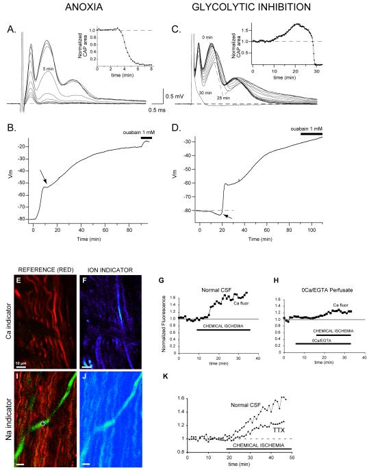
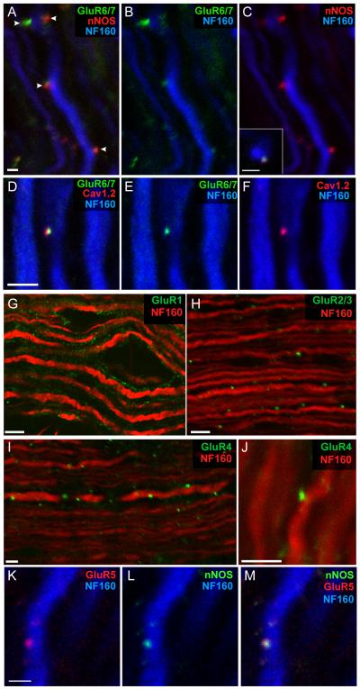
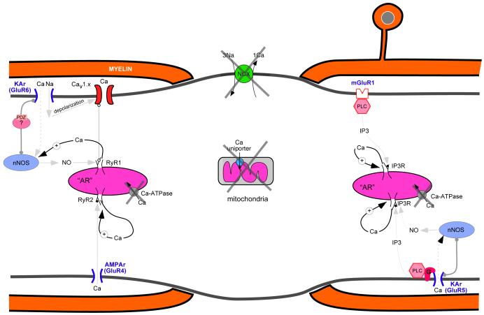
Axonal degeneration causes morbidity in many neurological conditions including stroke, neurotrauma and multiple sclerosis. The limited ability of central nervous system (CNS) neurons to regenerate, combined with the observation that axonal damage causes clinical disability, has spurred efforts to investigate the mechanisms of axonal degeneration. Ca influx from outside the axon is a key mediator of injury. More recently, substantial pools of intra-axonal Ca sequestered in the 'axoplasmic reticulum' have been reported. These Ca stores are under the control of multimolecular 'nanocomplexes' located along the internodes under the myelin. The overactivation of these complexes during disease can lead to a lethal release of Ca from intra-axonal stores. Rich receptor pharmacology offers tantalizing therapeutic options targeting these nanocomplexes in the many diseases where axonal degeneration is prominent.


Figures

Figure 1
Figure 1
Effect of anoxia or glycolytic block on optic nerve propagated compound action potentials (CAP) or resting membrane potentials (Vm). A: representative tracings show a rapid loss of the compound action potential recorded from rat optic nerve in vitro. Traces are displayed at 1 min intervals with anoxia beginning at time zero. Inset shows rate of decline of the area under the CAP. B: graph of rat optic nerve compound Vm recorded in a grease gap chamber. Chemical anoxia (2 mM NaCN) induces a rapid membrane depolarization that coincides with the loss of excitability as shown in panel A. After 10–15 min of anoxia there is an abrupt change in the rate of depolarization (arrow), which is never seen with ouabain application alone. This might reflect intrinsic mechanisms designed to limit a potentially deleterious membrane depolarization. Subsequent inhibition of Na+-K+-ATPase with ouabain after 90 min of anoxia induces an additional small depolarization indicating a minor contribution of glycolysis to Na+ and K+ pumping. C: in contrast to anoxia, the effects of glycolytic inhibition (1 mM iodoacetate) are delayed 10–20 min. CAP tracings are shown at intervals of 2 min. The initial manifestation is a rise in the CAP area that can exceed control area by 50% or more, as shown in the inset. A delay in peak latencies also occurs (compare tracings from time zero and 28 min); taken together, these features are characteristic of a transient initial hyperpolarization (see panel D). Excitability then fails very abruptly. D: Vm during glycolytic inhibition illustrates the delayed effects, with the initial change being a transient hyperpolarization (arrow) which closely coincides with the increase in CAP area illustrated in panel C. This is then followed by a rapid and massive depolarization corresponding to the sudden loss of excitability (panel C). Application of ouabain after 90 min of glycolytic block has no effect, indicating complete Na+-K+-ATPase failure under these conditions (compare with panel B). In parallel with electrophysiological failure, there is accumulation of abnormal levels of Ca (E-H) and Na (I-K) ion in ischemic axons, outlined by a red reference dye in E and I (the asterisk denotes a dye-filled capillary). The Ca and Na fluorescent indicators are shown in pseudocolor (F, J). Axonal Ca increases even in the absence of extracellular Ca (H). The Na rise is significantly, but incompletely, inhibited by blocking voltage-gated Na channels with tetrodotoxin (TTX) (K). Modified from refs [15, 17] with permission.
Figure 2
Figure 2
Sources of intra-axonal Ca release. A. Central myelinated axons possess formidable intracellular stores of Ca containing sufficient quantities to cause significant damage to the fiber. The Ca pools are stored in the axoplasmic reticulum (AR), the axonal analog of the endoplasmic reticulum (ER) in other cells, and mitochondria. The main intracellular Ca release channels are ryanodine receptors [1] (normally activated by Ca or by depolarization sensed by axolemmal voltage-sensitive Ca channels) and channels stimulated by inositol trisphosphate Ins(1,4,5)P3 [2]. Na loading of the axon through nodal Nav1.6 promotes Ca release from mitochondria by reverse mitochondrial Na-Ca exchange [3]. B. Proposed relationship between axonal glutamate receptors and intra-axonal Ca stores. The intracellular Ca release channels are under complex control of neurotransmitter receptors (mainly glutamate), expressed in clusters, together with related signaling molecules, along the internodal axolemma under the myelin sheath. GluR4 AMPA receptors permeate small amounts of Ca that, in turn, release Ca from the AR via ‘cardiac-type’ Ca-induced Ca release [1]. By contrast, axonal Ca increases from activation of GluR5 kainate receptors occur mainly via a G-protein-coupled, phospholipase C (‘PLC’)-dependent synthesis of IP3, which in turn activates Ins(1,4,5)P3 receptors on the AR [2]; this latter mechanism is partially dependent on NO, which is synthesized by neuronal nNOS, itself activated by small amounts of Ca entry via the GluR5 receptor; the locally produced NO may then further upregulate Ins(1,4,5)P3 receptor activity. Group I metabotropic glutamate receptors also activate Ins(1,4,5)P3-dependent Ca stores via PLC [3]. Stimulation of GluR6 kainate receptors induces a local depolarization and a small amount of Ca entry. The depolarization activates L-type Ca channels (Cav), whereas the kainate-receptor-mediated Ca2+ influx stimulates nNOS, which is scaffolded in the vicinity of the receptor. A PDZ-containing adaptor protein likely plays a role in organizing at least some of the signaling molecules into a functional ‘nanocomplex’ [4]. As with GluR5 receptors, locally generated NO might upregulate the activity of ryanodine receptors, which are activated by the depolarization-induced conformational change of the Ca channel, leading to release of Ca from the AR. The physiological roles of these nanocomplexes are unknown. However, overactivation of these signaling molecules by pathological levels of glutamate, NO and depolarization could be a major determinant of axonal degeneration in a variety of disorders. Mitochondria are omitted from panel B for clarity. xx[dhh5]
Figure 3
Figure 3
The internodal axolemma of myelinated CNS fibers expresses a rich variety of glutamate receptors and related signaling molecules. (A–C) Triple-immunolabeled spinal axons exhibit punctate regions of co-localized GluR6/7 and nNOS clusters (arrowheads) at the surface of neurofilament-stained axon cylinders. Inset: transverse view of a surface cluster in another fiber. (D–F) GluR6/7 clusters also co-localize with Cav1.2 L-type Ca2+ channels. AMPA receptor subunits are also expressed on myelinated axons: (G) GluR1 staining was patchy but is seen mainly in the myelin sheath. (H–J) By contrast, GluR2/3 (mainly GluR3 given the absence of GluR2 in myelinated axons) and GluR4 staining was punctate, and was localized at the surface of neurofilament-positive axonal profiles. (K–M) Likewise, GluR5 kainate receptor subunits were also expressed on the surfaces of axons in punctate areas, co-localized with nNOS. Scale bars: A–F, K–M, 2 μm; G–J 5 μm. Modified from refs. [54, 55] with permission. xx[dhh6]
Figure 4
Figure 4
Potential positive feedback of internal Ca release leading to a runaway emptying of intra-axonal stores. Under normal conditions, Ca homeostatic mechanisms (membrane Na-Ca exchanger [NCX], axoplasmic reticulum Ca-ATPase, Ca-binding proteins, mitochondria) maintain tight control over cellular Ca fluctuations. During pathological states, however, failure of Ca homeostasis (large Xs depict failure of the major Ca homeostatic systems) not only leads to excessive accumulation of released Ca, but this released Ca may in turn promote accelerated and unrestrained additional release of this ion by virtue of the Ca-dependence of nNOS, phospholipase C, and intracellular ryanodine and IP3 Ca channels. This may ultimately cause an irreversible and catastrophic accumulation of axonal Ca levels, causing structural and functional demise of the axon.

Similar articles

Cited by

References

    1. Waxman SG, Kocsis JD, Stys PK. The Axon : structure, function and pathophysiology. Oxford University Press; New York; Oxford: 1995. p. xv.p. 692. [2] of col. plates.
    1. Wolf JA, et al. Traumatic axonal injury induces calcium influx modulated by tetrodotoxin-sensitive sodium channels. J Neurosci. 2001;21(6):1923–30. - PMC - PubMed
    1. Trapp BD, Stys PK. Virtual hypoxia and chronic necrosis of demyelinated axons in multiple sclerosis. Lancet Neurol. 2009;8(3):280–91. - PubMed
    1. Stys PK. General mechanisms of axonal damage and its prevention. J Neurol Sci. 2005;233(1-2):3–13. - PubMed
    1. Wilkins A, Scolding N. Protecting axons in multiple sclerosis. Mult Scler. 2008;14(8):1013–25. - PubMed

Publication types

MeSH terms