. 2010 May 7;285(19):14285-91.
doi: 10.1074/jbc.M109.087023. Epub 2010 Mar 5.

Mechanistic insight into the function of the C-terminal PKD domain of the collagenolytic serine protease deseasin MCP-01 from deep sea Pseudoalteromonas sp. SM9913: binding of the PKD domain to collagen results in collagen swelling but does not unwind the collagen triple helix

Affiliations

Mechanistic insight into the function of the C-terminal PKD domain of the collagenolytic serine protease deseasin MCP-01 from deep sea Pseudoalteromonas sp. SM9913: binding of the PKD domain to collagen results in collagen swelling but does not unwind the collagen triple helix

Yu-Kai Wang et al. J Biol Chem. .

Abstract

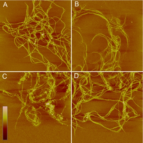
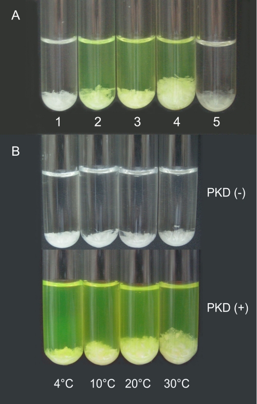
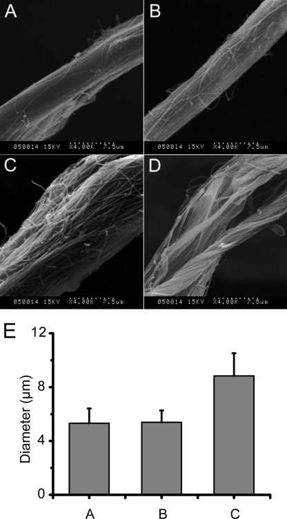
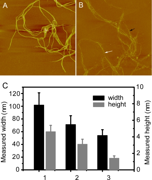
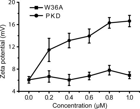
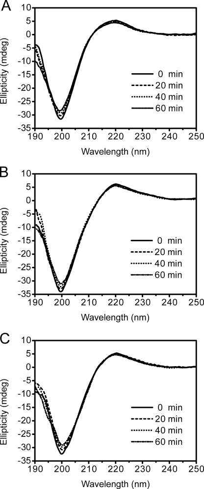
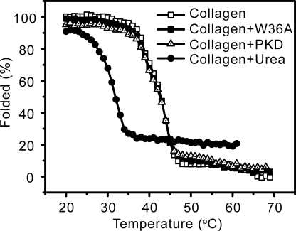
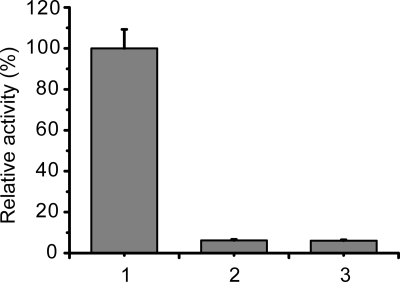
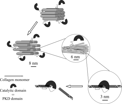
Deseasin MCP-01 is a bacterial collagenolytic serine protease. Its catalytic domain alone can degrade collagen, and its C-terminal PKD domain is a collagen-binding domain (CBD) that can improve the collagenolytic efficiency of the catalytic domain by an unknown mechanism. Here, scanning electron microscopy (SEM), atomic force microscopy (AFM), zeta potential, and circular dichroism spectroscopy were used to clarify the functional mechanism of the PKD domain in MCP-01 collagenolysis. The PKD domain observably swelled insoluble collagen. Its collagen-swelling ability and its improvement to the collagenolysis of the catalytic domain are both temperature-dependent. SEM observation showed the PKD domain swelled collagen fascicles with an increase of their diameter from 5.3 mum to 8.8 mum after 1 h of treatment, and the fibrils forming the fascicles were dispersed. AFM observation directly showed that the PKD domain bound collagen, swelled the microfibrils, and exposed the monomers. The PKD mutant W36A neither bound collagen nor disturbed its structure. Zeta potential results demonstrated that PKD treatment increased the net positive charges of the collagen surface. PKD treatment caused no change in the content or the thermostability of the collagen triple helix. Furthermore, the PKD-treated collagen could not be degraded by gelatinase. Therefore, though the triple helix monomers were exposed, the PKD domain could not unwind the collagen triple helix. Our study reveals the functional mechanism of the PKD domain of the collagenolytic serine protease MCP-01 in collagen degradation, which is distinct from that of the CBDs of mammalian matrix metalloproteases.


Figures

FIGURE 1.
FIGURE 1.
AFM observation of the binding of the PKD domain to collagen. A, self-assembled collagen in 50 mm Tris-HCl (pH 8.5) incubated at 30 °C for 1 h. B, collagen incubated with EGFP at 30 °C for 1 h. C, collagen incubated with EGFP-PKD at 30 °C for 1 h. D, collagen incubated with EGFP-W36A at 30 °C for 1 h. Scale bar: 30 nm. Scan size: A, 6 μm; B–D, 5 μm.
FIGURE 2.
FIGURE 2.
The collagen-swelling effect of the PKD domain. A, collagen swollen by the PKD domain or urea at 20 °C for 1 h. A total of 10 mg of insoluble type I collagen was incubated in 2 ml of 20 mm borate buffer (pH 8.5) (1), 20 mm borate buffer containing 6 μm EGFP (2), 20 mm borate buffer containing 6 μm EGFP-W36A (3), 20 mm borate buffer containing 6 μm EGFP-PKD (4), or 20 mm borate buffer containing 6 m urea (5). B, collagen swollen by the PKD domain for 1 h at different temperatures. A total of 10 mg of insoluble type I collagen in 2 ml of 20 mm borate buffer (pH 8.5) was incubated with 6 μm EGFP-PKD at 4, 10, 20, and 30 °C for 1 h each.
FIGURE 3.
FIGURE 3.
SEM observation of collagen fascicles swollen by the PKD domain. A total of 10 mg of type I insoluble collagen in 1 ml of 20 mm borate buffer (pH 8.5) was incubated at 20 °C with 3 μm EGFP-PKD or EGFP-W36A. The samples were observed using scanning electron microscopy (Hitachi S-570) by Usha and Ramasami's method (23). A, collagen fascicle incubated at 20 °C for 1 h. B, collagen fascicle incubated with W36A-EGFP at 20 °C for 1 h. C, collagen fascicle incubated with EGFP-PKD at 20 °C for 0.5 h. D, collagen fascicle incubated with EGFP-PKD at 20 °C for 1 h. E, diameter of untreated and EGFP-W36A- and EGFP-PKD-treated collagen fascicles. Columns in E show the diameter of: 1) untreated collagen fascicles, 2) EGFP-W36A-treated collagen fascicles, and 3) EGFP-PKD-treated collagen fascicles. All data in E are averages of the data from 50 collagen fascicles measured.
FIGURE 4.
FIGURE 4.
A, AFM micrograph of collagen microfibrils incubated at 30 °C for 1 h. B, AFM micrograph of collagen microfibrils incubated with EGFP-PKD at 30 °C for 1 h. The black arrow shows the relatively thicker collagen microfibrils, and the white arrow shows the relatively thinner collagen microfibrils after incubation with EGFP-PKD. Scan size is 5 μm in both A and B. C, height and width of the PKD-untreated and PKD-treated collagen microfibrils calculated from AFM micrographs A and B. Columns in C show the height and width of: 1) the PKD-untreated collagen microfibrils in AFM micrograph A, 2) the thicker collagen microfibrils in AFM micrograph B, and 3) the thinner collagen microfibrils in AFM micrograph B. All data in C are averages of the data from 20 sites measured.
FIGURE 5.
FIGURE 5.
Zeta potential of collagen treated with different concentrations of EGFP-PKD or EGFP-W36A at 20 °C for 1 h.
FIGURE 6.
FIGURE 6.
CD spectra of collagen at 20 °C for 1 h (A), collagen incubated with EGFP-PKD at 20 °C for 1 h (B), and collagen incubated with EGFP-W36A (C) at 20 °C for 1 h.
FIGURE 7.
FIGURE 7.
Thermal unfolding curves of untreated collagen and collagens treated with EGFP-PKD, EGFP-W36A, or urea at 20 °C for 1 h.
FIGURE 8.
FIGURE 8.
Activities of chymotrypsin against thermally denatured collagen (1), PKD-untreated collagen (2), and PKD-treated collagen (3). Thermally denatured collagen was prepared by heating insoluble type I collagen at 65 °C for 20 min. PKD-treated collagen was prepared by incubating insoluble type I collagen with 3 μm PKD at 20 °C for 1 h. Chymotrypsin activity was measured at 37 °C by a previously described method (10).
FIGURE 9.
FIGURE 9.
A schematic diagram of the mechanism of collagenolysis by serine protease MCP-01. During collagenolysis by MCP-01, the C-terminal PKD domain binds collagen, swells collagen aggregate structures, fascicles, fibrils, and microfibrils, and finally exposes collagen triple-helical monomers. Then, the N-terminal catalytic domain accommodates the triple-helical collagen monomer in its catalytic cavity and cleaves it.

Similar articles

Cited by

References

    1. Gluecksmann-Kuis M. A., Tayber O., Woolf E. A., Bougueleret L., Deng N., Alperin G. D., Iris F., Hawkins F. (1995) Cell 81, 289–298 - PubMed
    1. Bycroft M., Bateman A., Clarke J., Hamill S. J., Sandford R., Thomas R. L., Chothia C. (1999) EMBO J. 18, 297–305 - PMC - PubMed
    1. Jing H., Takagi J., Liu J. H., Lindgren S., Zhang R. G., Joachimiak A., Wang J. H., Springer T. A. (2002) Structure 10, 1453–1464 - PubMed
    1. Perrakis A., Tews I., Dauter Z., Oppenheim A. B., Chet I., Wilson K. S., Vorgias C. E. (1994) Structure 2, 1169–1180 - PubMed
    1. Orikoshi H., Nakayama S., Hanato C., Miyamoto K., Tsujibo H. (2005) J. Appl. Microbiol. 99, 551–557 - PubMed

Publication types